plugin

Description

SIMPLETRACKER a simple particle tracking algorithm that can deal with gaps.

Tracking , or particle linking, consist in re-building the trajectories of one or several particles as they move along time. Their position is reported at each frame, but their identity is yet unknown: we do not know what particle in one frame corresponding to a particle in the previous frame. Tracking algorithms aim at providing a solution for this problem. 

simpletracker.m is - as the name says - a simple implementation of a tracking algorithm, that can deal with gaps. A gap happens when one particle that was detected in one frame is not detected in the subsequent one. If not dealt with, this generates a track break, or a gap, in the frame where the particle disappear, and a false new track in the frame where it re-appear. 

need a thumbnail
Description

Mean square displacement (MSD) analysis is a technique commonly used in colloidal studies and biophysics to determine what is the mode of displacement of particles followed over time. In particular, it can help determine whether the particle is:

  • freely diffusing;
  • transported;
  • bound and limited in its movement.

On top of this, it can also derive an estimate of the parameters of the movement, such as the diffusion coefficient.

@msdanalyzer is a MATLAB per-value class that helps performing this kind of analysis. The user provides several trajectories he measured, and the class can derive meaningful quantities for the determination of the movement modality, assuming that all particles follow the same movement model and sample the same environment.

has function
Examples of tracks to perform MSD analysis.
Description

R wrapper around the OMERO Java Gateway, to enable access to OMERO via R using rJava

has function
need a thumbnail
Description

ScientiFig is a free tool to help you create, format or reformat scientific figures. It comes either as a stand alonesoftware, either as a Fiji/IJ plugin.

has topic
has function
scientifig
Description

This one example workflow from the Cell Profiler(CP)  Examples . CP is commonly used to count cells or other objects as well as percent-positives, by measuring the per-cell staining intensity. This pipeline shows how to do both of these tasks, and demonstrates how various modules may be used to accomplish the same result. 

In a few words, it used the IdentifyPrimaryObject module of CellProfiler to detect nuclei from a channel (e.g DAPI), then again the same module on another channel to detect another probe (e.g some particular histone)  .

Then objects (nuclei) are related to the second object (Histone), to create a parent child-relation ship: where nuclei can have histone has child. Nuclei are then filtered according to the property of having histone (positive) or not having histone (negtiveobject) related to them.  If needed, nuclei can be expanded in order to include touching object rather than object inside only.

The percentage of positive nuclei vs total number of nuclei can then be computed using the CalculateMath Module.

Positivepercentcell
Description

This workflow can be ran with data from 3D-SIM showing the centrosomes in order to compare the distribution of diameters of rings (or toroids) of different proteins from the centrioles or the peri centriolar material. It aims to reproduce the results of the Nature Cell Biology Paper Subdiffraction imaging of centrosomes reveals higher-order organizational features of pericentriolar material  from the same data set but with a different analysis method.

It is slightly different from the methods described in the paper itself, where the method was to work on a maximum intensity projection of a 3D-SIM stack, and then to fit circle to the centrioles to estimate the diameters of the toroids.

In this workflow, the images are read from the IDR , then process by thresholding (Maximum entropy auto thresholding with Image J), and processed by Analyze Particles  with different measurement sets, including the bouding box. Then the analysis of diameters and the statistical test are performed using R. All the code and data sets are available, and in the case of this paper have shown a layered organisation of the proteins.

Combined view from Figure 1 Lawo et al.
Description

A collection of components for super resolution image data:

  • Detect Molecules
  • Reconstruct Image
  • Results table
  • Drift correction
  • Chromatic correction
Description

This component can be used to find moving foreground features, which can be a powerful way to suppress false background detections in subsequent tracking steps.

set time window, and standard deviations above background for foreground time window should be more than 2x larger than time taken for a feature to traverse a pixel (NB. total window is 2x half-width +1) moving foreground identified by intensity increase relative to background average (i.e. median) for a pixel over a given time window "soft" segmentation, yielding foreground probability related to excess intensity (in standard deviations) over background level crude Anscombe transform applied to data to stabilize the variance

need a thumbnail
Description

This is a plugin bundled with native ImageJ.

See IJ reference for more details > Link

need a thumbnail
Description

Manual tracking using Trackmate plugin (comes with FIji, so no installation required if you are using Fiji). 

has function
Description

The freely available software module below is a 3D LoG filter. It applies a LoG (Laplacian of Gaussian or Mexican Hat) filter to a 2D image or to 3D volume. Here, we have a fast implementation. It is a perfect tool to enhance spots, like spherical particles, in noisy images. This module is easy to tune, only by selecting the standard deviations in X, Y and Z directions.

IJ Macro command example
run("LoG 3D", "sigmax=1 sigmay=1 sigmaz=13 displaykernel=0 volume=1");
Description

This method was originally designed to track objects (not necessarily spots) already identified in 2D 
frames and has been applied previously to particle tracking and analysis in high-speed atomic force microscopy image series.


need a thumbnail
Description


The phase contrast microscopy segmentation toolbox (PHANTAST) is a collection of open-source algorithms and tools for the processing of phase contrast microscopy (PCM) images. It was developed at University College London's department of Biochemical Engineering and CoMPLEX.

has function
Description

An ImageJ plugin for DEFCoN, the fluorescence spot counter based on fully convolutional neural networks

has topic
Description

Align_slices in stack utilized the template matching function cvMatch_Template to do slice registration(alignment) based on a selected landmark.
This function will try to find the landmark or the most similar image pattern in every slice and translate each slice so that the landmark pattern will be the same position throughout the whole stack. It could be used to fix the drift of a time-lapse image stacks.

Source code: link

Input data: image stack
output data: image stack

has function
Description

It implements the template matching function from the OpenCV library. The java interface of OpenCV was done through the javacv library. It is quite similar as the existing template matching plugin but runs much faster and users could choose among six matching methods: 

1.Squared difference

2.Normalized squared difference

3.Cross-correlation

4.Normalized cross-correlation

5.Correlation coefficient

6.Normalized correlation coefficient

The detailed algorithms could be found here.

The cvMatch_Template will search a specific object (image pattern) over an image of interest by the user-specified method. 

Description

This ImageJ plugin contains two functions. The first one is the cvMatch_Template. It implements the template matching function from the OpenCV library. The second function Align_slices in stack utilized the previous matching function to do slice registration(alignment) based on a selected landmark. 

For more details, refer to the page of each component. 

cvMatch_Template

Align Slices in Stack

has function
Description
HyphaTrackerWorkflow
HyphaTracker Workflow

HyphaTracker propose a workflow for time-resolved analysis of conidia germination. Each part of this workflow can also be used independnatly , as a toolbox. It has been tested on bright-field microscopic images of conidial germination. Its purpose is mainly to identify the germlings and to remove crossing hyphae, and measure the dynamics of their growth.

hyphatracker
Description

LimeSeg: A coarsed-grained lipid membrane simulation for 3D image segmentation

Download instruction:

There is no download but you can easily install this plugin via ImageJ update site. If you reallu need to download the jar file, access the file in the update site repository (Link)

has function
Description

automated open-source image acquisition and on-the-fly analysis pipeline (initially developped for analysis of mitotic defects in fission yeast)

maars workflow from publication


maars

CEM

Description

Computer-assisted Evaluation of Myelin formation (CEM) is a collection designed to automate myelin quantification. It requires use input to choose the best threshold values. The myelin is calculated as an overlap between neuronal signal and oligodendrocyte signal. Results are given as pixel counts and percents.

CEM runs as an imageJ plugin with an optional Matlab extension to remove cell bodies. More details are published at Kerman et al. 2015 Development. Supplemental Material includes a detailed user manual and the download link.

Myelin
Description

MaMuT is an end user plugin that combines the BigDataViewer and TrackMate to provide an application that allow browsing, annotating and curating annotations for large image data.

Description

Kymograph generation under ImageJ:

one simple solution, plot a line (ROI line) on the first frame, where you want to generate the kymograph.

Use

Image  / Stacks  / Reslice

It will generate a new image were Y dimension is the time, and X the position on the line you have drawn.

need a thumbnail
Description

  FlyLimbTracker is  a method that uses active contours to semi-automatically track body and leg segments from video image sequences of unmarked, freely behaving Drosophila flies. This approach can be used to measure leg segment motions during a variety of locomotor and grooming behaviors.

For now the plugin have to be downlaoded directly from the EPFL website (see link), not from the search bar as usual in ICY.


Drosophila track legs
Description

Working version of a simple GUI frontend for CMTK image registration tools in Fiji

need a thumbnail
Description

Estimate the positions and spacing between sections (or at local points) of three dimensional image data. This method may be applied to any imaging modality that acquires 3-dimensional data as a stack of 2-dimensional sections. We provide plugins for both Fiji and TrakEM2.

has function
Description

In light-sheet microscopy, overall image content and resolution are improved by acquiring and fusing multiple views of the sample from different directions. State-of-the-art multi-view (MV) deconvolution simultaneously fuses and deconvolves the images in 3D, but processing takes a multiple of the acquisition time and constitutes the bottleneck in the imaging pipeline. Here, we show that MV deconvolution in 3D can finally be achieved in real-time by processing cross-sectional planes individually on the massively parallel architecture of a graphics processing unit (GPU). Our approximation is valid in the typical case where the rotation axis lies in the imaging plane.

need a thumbnail
Description

This R package implements the NBLAST neuron similarity algorithm described in a preprint available at http://dx.doi.org/10.1101/006346. In addition to basic pairwise comparison, the package implements search of databases of neurons. There is also suport for all x all comparison for a group of neurons. This can produce a distance matrix suitable for hierarchical clustering, which is also implemented in the package.

has topic
has function
Description

NeuroGPS-Tree is a workflow developed to reconstruct a neuronal population from a dense, large-scale data set. NeuroGPS-Tree is suitable for processing image stacks acquired by different image modalities.

need a thumbnail
Description

MTrack is a tool, which detects, tracks, and measures the behavior of fluorescently labeled microtubules imaged by TIRF (total internal reflection fluorescence) microscopy. In such an in vitro reconstitution approach, stabilized, non-dynamic microtubule seeds serve as nucleation points for dynamically growing microtubules.

MTrack is a bi-modular tool. The first module detects and tracks the growing microtubule ends and creates trajectories. The second module uses these trajectories to fit models of dynamic behavior (polymerization and depolymerization velocities, catastrophe and rescue frequencies). It also computes statistics such as length and lifetime distributions when analyzing more than one movie (batch mode).

has topic
Track Filament shaped objects and analyze tracks using Ransac fits.
Description

Calculates and corrects for lens-distortion models including chromatic abberation from confocal stacks.

Description

SliceMap

Whole brain tissue slices are commonly used in neurobiological research for analyzing pathological features in an anatomically defined manner. However, since many pathologies are expressed in specific regions of the brain, it is necessary to have an annotation of the regions in the brain slices. Such an annotation can be done by manual delineation, as done most often, or by an automated region annotation tool.

SliceMap is a FIJI/ImageJ plugin for automated brain region annotation of fluorescent brain slices. The plugin uses a reference library of pre-annotated brain slices (the brain region templates) to annotate brain regions of unknown samples. To perform the region annotation, SliceMap registers the reference slices to the sample slice (using elastic registration plugin BUnwarpJ) and uses the resulting image transformations to morph the template regions towards the anatomical brain regions of the sample. The resulting brain regions are saved as FIJI/ImageJ ROI’s (Regions Of Interest) as a single zip-file for each sample slice.

More information can also be found in "SliceMap: an algorithm for automated brain region annotation", Michaël Barbier, Astrid Bottelbergs, Rony Nuydens, Andreas Ebneth, Winnok H De Vos, Bioinformatics, btx658, https://doi.org/10.1093/bioinformatics/btx658

Example: SliceMaps brain region segmentation
Description

The Multiview Reconstruction software package enables users to register, fuse, deconvolve and view multiview microscopy images. The software is designed for lightsheet fluorescence microscopy (LSFM), but is applicable to any form of three or higher dimensional imaging modalities like confocal timeseries or multicolor stacks. 

need a thumbnail
Description

The BigDataViewer is a re-slicing browser for terabyte-sized multi-view image sequences. BigDataViewer was developed with multi-view light-sheet microscopy data in mind and integrates well with Fiji's SPIMage processing pipeline.

Description

Bigwarp is a tool for manual, interactive, landmark-based deformable image alignment. It uses the BigDataViewer for visualization and navigation, and uses a Thin Plate Spline implemented in Java to build a deformation from point correspondences.

Bigwarp screenshot
Description

The BigStitcher is a software package that allows simple and efficient alignment of multi-tile and multi-angle image datasets, for example acquired by lightsheet, widefield or confocal microscopes. The software supports images of almost arbitrary size ranging from very small images up to volumes in the range of many terabytes, which are for example produced when acquiring cleared tissue samples with lightsheet microscopy.

Description

FracLac is for digital image analysis. Use it to measure difficult to describe morphological features.
FracLac is a plugin for ImageJ. It is freely available software developed and maintained by our lab at the School of Community Health, Faculty of Science, Charles Sturt University, Australia. The author of the software and project lead is also the author of this document (me, Audrey Karperien). The basic box counting algorithm was originally modified from ImageJ's box counting algorithm and H. Jelinek's NIH Image plugin, and was further elaborated based on extensive research and development. The convex hull algorithm was provided by Thomas Roy, University of Alberta, Canada. As open source software, with the continuing help of a host of users and collaborators, FracLac has evolved to a suite of fractal analysis and morphology functions.

need a thumbnail
Description

Quote: "The GDSC ImageJ plugins are a collection of analysis programs for microscopy images including colocalisation analysis and peak finding (FindFoci)."

Many types of analysis besides simply finding foci detection (spot detection) is bundled in this plugin. One prominent function is "FindFoci Optimizer". This allows feeding images with spot annotation by the user (multi-point selection tool) and scans through various parameter combinations to find the best parameter set that gives the results similar to the annotation. This is almost like machine learning... but with well-established parameter types that allows you to fully understand what is going on.

Description

This plugin filters a 3D image stack (or 2D image) to produce a score for how "tube-like" each point in the image is. This is useful as a preprocessing step for tracing neurons or blood vessels, for example. For 3D image stacks, the plugin uses the eigenvalues of the Hessian matrix to calculate this measure of "tubeness", using a metrics mentioned in Sato et al 1997 ¹: if the larger two eigenvalues (λ₂ and λ₃) are both negative then value is √(λ₂λ₃), otherwise the value is 0. For 2D images, if the large eigenvalue is negative, we return its absolute value and otherwise return 0.

This plugin is now bundled as part of Fiji.

Description

The invention comprises a software tool, NeuronMetrics, which functions as a set of modules that run in the open-source program ImageJ. NeuronMetrics features a novel method for estimating neural “branch number” (a measure of the axonal complexity) from two-dimensional images. In addition, the tool features a novel method for modeling neural structure in large “gaps” that result from image artifacts.


has topic
need a thumbnail
Description

This plugin achieves easy creation of image figures for publications, reports, projects.

  • Easy-to-design interactive figure layout.

  • Visually assign image content to panels.

  • High-quality image scaling and rotation.

  • Easy and consistent panel labels and scale bars.

  • Each panel has it's original datasource's properties and tracks achieved image processing.

  • Save and re-open editable figures.

  • Export as standard image formats with textual description of each panel history.

Compared to Make montage, the plugin adds more flexibility to montage creation: Easy-to-design interactive figure layout. Visually assign image content to panels. High-quality image scaling and rotation. Easy and consistent panel labels and scale bars. Each panel has it's original data source's properties and tracks achieved image processing. Save and re-open editable figures. Export as standard image formats with textual description of each panel history. 

has topic
has function
FigureJ
Description

hIPNAT (hIPNAT: Image Processing for NeuroAnatomy and Tree-like structures) is a set of tools for the analysis of images of neurons and other tree-like morphologies. It is written for ImageJ, the de facto standard in scientific image processing. It is available through the ImageJ Neuroanatomy update site.

need a thumbnail
Description

"we present a new fully automated 3D reconstruction algorithm, called TReMAP, short for Tracing, Reverse Mapping and Assembling of 2D Projections. Instead of tracing a 3D image directly in the 3D space as seen in majority of the tracing methods, we first trace the 2D projection trees in 2Dplanes, followed by reverse-mapping the resulting 2D tracing results back into the 3D space as 3D curves; then we use a minimal spanning tree (MST) method to assemble all the 3D curves to generate the final 3D reconstruction. Because we simplify a 3D reconstruction problem into 2D, the computational costs are reduced dramatically." 

Suitable for high throughput neuron image analysis (image sizes >10GB). This plugin can be used with default parameters or user-defined parameters.

Example_TReMAP_Result
Description

"We have developed an automatic graph algorithm, called the all-path pruning (APP), to trace the 3D structure of a neuron. To avoid potential mis-tracing of some parts of a neuron, an APP first produces an initial over-reconstruction, by tracing the optimal geodesic shortest path from the seed location to every possible destination voxel/pixel location in the image. Since the initial reconstruction contains all the possible paths and thus could contain redundant structural components (SC), we simplify the entire reconstruction without compromising its connectedness by pruning the redundant structural elements, using a new maximal- covering minimal-redundant (MCMR) subgraph algorithm. We show that MCMR has a linear computational complexity and will converge. We examined the performance of our method using challenging 3D neuronal image datasets of model organisms (e.g. fruit fly)"

This plugin can be used with default parameters or user-defined parameters.

APP_Vaa3D_example_results
Description

Summary

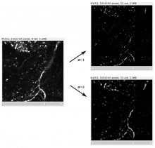
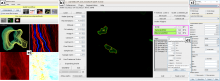
QuimP is software for tracking cellular shape changes and dynamic distributions of fluorescent reporters at the cell membrane. QuimP's unique selling point is the possibility to aggregate data from many cells in form of spatio-temporal maps of dynamic events, independently of cell size and shape. QuimP has been successfully applied to address a wide range of problems related to cell movement in many different cell types. 

Introduction

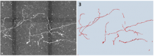
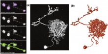
In transmembrane signalling the cell membrane plays a fundamental role in localising intracellular signalling components to specific sites of action, for example to reorganise the actomyosin cortex during cell polarisation and locomotion. The localisation of different components can be directly or indirectly visualised using fluorescence microscopy, for high-throughput screening commonly in 2D. A quantitative understanding demands segmentation and tracking of whole cells and fluorescence signals associated with the moving cell boundary, for example those associated with actin polymerisation at the cell front of locomoting cells. As regards segmentation, a wide range of methods can be used (threshold based, region growing, active contours or level sets) to obtain closed cell contours, which then are used to sample fluorescence adjacent to the cell edge in a straightforward manner. The most critical step however is cell edge tracking, which links points on contours at time t to corresponding points at t+1. Optical flow methods have been employed, but usually fail to meet the requirement that total fluorescence must not change. QuimP uses a method (ECMM, electrostatic contour migration method (Tyson et al., 2010) which has been shown to outperform traditional level set methods. ECMM minimises the sum of path lengths connecting all pairs of points, equivalent to minimising the energy required for cell deformation. The original segmentation based on an active contour method and outline tracking algorithms have been described in (Dormann et al., 2002; Tyson et al., 2010; Tyson et al., 2014).

Screenshot
Description

SRRF is a high-performance analytical approach for Live-cell Super-Resolution Microscopy, provided as a fast GPU-enabled ImageJ plugin. SRRF is capable of extracting high-fidelity super-resolution information from TIRF, widefield and confocals using conventional fluorophores such as GFP. SRRF is capable of live-cell imaging over timescales ranging from minutes to hours.

Comparison TIRF - SRRF