Single molecule localisation

Synonyms
PAINT image reconstruction
STORM image reconstruction
SMLM image reconstruction
Single-molecule localisation microscopy image reconstruction
PALM image reconstruction
Description

MATLAB app to characterize nanoparticles imaged with super-resolution microscopy. nanoFeatures will read text and csv files from the NIKON and ONI microscopes and from the ThunderSTORM Fiji plugin, then cluster the localizations and filter by size and sphericity and finally output nanoparticle features like size, aspect ratio, and number of localizations per cluster (total and for each channel).

GUI first tab to browse and input files, select input type and check extra filters if needed.
Description

The PYthon Microscopy Environment is an open-source package providing image acquisition and data analysis functionality for a number of microscopy applications, but with a particular emphasis on single molecule localisation microscopy (PALM/STORM/PAINT etc ...). The package is multi platform, running on Windows, Linux, and OSX.

It comes with 3 main modules:

  • PYMEAcquire - Instrument control and simulation
  • dh5view - Image Data Analysis and Viewing
  • VisGUI - Visualising Localization Data Sets
Description

This script includes a rough feature detection and then fine 2D Gaussian algorithm to fit Gaussians within detected regions. This macro is unique because the ImageJ/Fiji curve fitting API only supports 1-D curve. I get around this by linearising the equation. This implementation is for isotropic (spherical) or anistropic (longer in x/y) diagonally covariant Gaussians but not fully covariant Gaussians (anisotropic and rotated). 

Description

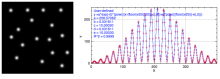
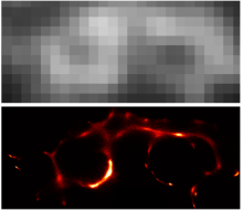
Stochastic optical reconstruction microscopy (STORM) and related methods achieves sub-diffraction-limit image resolution through sequential activation and localization of individual fluorophores. The analysis of image data from these methods has typically been confined to the sparse activation regime where the density of activated fluorophores is sufficiently low such that there is minimal overlap between the images of adjacent emitters. Recently several methods have been reported for analyzing higher density data, allowing partial overlap between adjacent emitters. However, these methods have so far been limited to two-dimensional imaging, in which the point spread function (PSF) of each emitter is assumed to be identical.

In this work, we present a method to analyze high-density super-resolution data in three dimensions, where the images of individual fluorophores not only overlap, but also have varying PSFs that depend on the z positions of the fluorophores.


need a thumbnail