Java

Description

The best way to start writing an ImageJ2 plugin (ImageJ2 developers call it command and not plugin) is to download the example command from github and modify it. There is a video tutorial on the whole workflow on how to do this on youtube.

has function
Description

AutoPilot is the open source project that hosts the general algorithm for fast and robust assessment of local image quality, an automated computational method for image-based mapping of the three-dimensional light-sheet geometry inside a fluorescently labeled biological specimen, and a general algorithm for data-driven optimization of the system state of light-sheet microscopes capable of multi-color imaging with multiple illumination and detection arms.

has function
Description

This plugin detects a minimum cost z-surface in a 3D volume. A z surface is a topographic map indicating the altitude z as a function of the position (x,y) in the image. The cost of the surface depends on pixel intensity the surface is going through. This plugin find the z-surface with the lowest intensity in an image.

has function
Description

The interactive Watershed Fiji plugin provides an interactive way to explore local maxima and threshold values while a resulting label map is updated on the fly.

After the user has found a reliable parameter configuration, it is possible to apply the same parameters to other images in a headless mode, for example via ImageJ macro scripting.

Description

Biocat is a java based software that allows to perform image classification or segmentation using machine learning. Several algorithm for the classification are available.

has topic
need a thumbnail
Description

SimpleITK provides a simplified interface to ITK in a variety of languages. A user can either download pre-built binaries, if they are available for the desired platform and language, or SimpleITK can be built from the source code. Currently, Python binaries are available on Microsoft Windows, GNU Linux and Mac OS X. C# and Java binaries are available for Windows. We are also working towards supporting R packaging.

need a thumbnail
Description

ZEN and APEER – Open Ecosystem for integrated Machine-Learning Workflows

Open ecosystem for integrated machine-learning workflows to train and use machine-learning models for image processing and image analysis inside the ZEN software or on the APEER cloud-based platform

Highlights ZEN

  • Simple User Interface for Labeling and Training
  • Engineered Features Sets and Deep Feature Extraction + Random Forrest for Semantic Segmentation
  • Object Classification workflows
  • Probability Thresholds and Conditional Random Fields
  • Import your own trained models as *.czann files (see: czmodel · PyPI)
  • Import "AIModel Containes" from arivis AI for advanced Instance Segmentation
  • Integration into ZEN Measurement Framework
  • Support for Multi-dimensional Datasets and Tile Images
  • open and standardized format to store trained models
ZEN Intellesis Segmentation

ZEN Intellesis Segmentation - Training UI

ZEN Intellesis - Pretrained Networks

ZEN Intellesis Segmentation - Use Deep Neural Networks

Intellesis Object Classification

ZEN Object Classification

Highlights Aarivis AI

  • Web-based tool to label datasets to train Deep Neural Networks
  • Fully automated hyper-parameter tuning
  • Export of trained models for semantic segmentation and AIModelContainer for Instance Segmentation
Annotation Tool

APEER Annotation Tool

Description

R wrapper around the OMERO Java Gateway, to enable access to OMERO via R using rJava

has function
need a thumbnail
Description

OpenImadis stands for Open Image Discovery: A platform for Image Life Cycle Management. It was previously called CID iManage (for Curie Image Database).

No image data conversions, no duplication.

- Uploads data to a secure server in the original format

- Unique id for data

Supports sharing and collaboration with access control

- Allows users to upload, view, update or download data based on their access privileges

Supports multiple ways of attaching meta-information

- Annotations, comments and file attachments

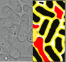
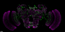
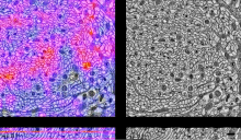
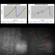
-Analysis results as query-able visual objects

Supports Archiving (data moving to another long-term storage but still searchable)

Facilitates custom visualization and analysis

- Access data from preferred analysis and visualization tools

- Access relevant bits of data to build efficient web and mobile application

Facilitate easy access to analysis and visualization applications hosted on other servers

- Run analysis on dedicated compute clusters

- Access applications hosted and published by other users

Highly Scalable

- Supports on-the-fly addition of server nodes to scale concurrent usage


openImadis
Description

A collection of components for super resolution image data:

  • Detect Molecules
  • Reconstruct Image
  • Results table
  • Drift correction
  • Chromatic correction
Description

This component can be used to find moving foreground features, which can be a powerful way to suppress false background detections in subsequent tracking steps.

set time window, and standard deviations above background for foreground time window should be more than 2x larger than time taken for a feature to traverse a pixel (NB. total window is 2x half-width +1) moving foreground identified by intensity increase relative to background average (i.e. median) for a pixel over a given time window "soft" segmentation, yielding foreground probability related to excess intensity (in standard deviations) over background level crude Anscombe transform applied to data to stabilize the variance

need a thumbnail
Description

This is a plugin bundled with native ImageJ.

See IJ reference for more details > Link

need a thumbnail
Description

Manual tracking using Trackmate plugin (comes with FIji, so no installation required if you are using Fiji). 

has function
Description

This method was originally designed to track objects (not necessarily spots) already identified in 2D 
frames and has been applied previously to particle tracking and analysis in high-speed atomic force microscopy image series.


need a thumbnail
Description

JAMA is a basic linear algebra package for Java. It provides user-level classes for constructing and manipulating real, dense matrices. It is meant to provide sufficient functionality for routine problems, packaged in a way that is natural and understandable to non-experts. It is intended to serve as the standard matrix class for Java, and will be proposed as such to the Java Grande Forum and then to Sun. A straightforward public-domain reference implementation has been developed by the MathWorks and NIST as a strawman for such a class. We are releasing this version in order to obtain public comment. There is no guarantee that future versions of JAMA will be compatible with this one.

need a thumbnail
Description


The phase contrast microscopy segmentation toolbox (PHANTAST) is a collection of open-source algorithms and tools for the processing of phase contrast microscopy (PCM) images. It was developed at University College London's department of Biochemical Engineering and CoMPLEX.

has function
Description

An ImageJ plugin for DEFCoN, the fluorescence spot counter based on fully convolutional neural networks

has topic
Description

Align_slices in stack utilized the template matching function cvMatch_Template to do slice registration(alignment) based on a selected landmark.
This function will try to find the landmark or the most similar image pattern in every slice and translate each slice so that the landmark pattern will be the same position throughout the whole stack. It could be used to fix the drift of a time-lapse image stacks.

Source code: link

Input data: image stack
output data: image stack

has function
Description

It implements the template matching function from the OpenCV library. The java interface of OpenCV was done through the javacv library. It is quite similar as the existing template matching plugin but runs much faster and users could choose among six matching methods: 

1.Squared difference

2.Normalized squared difference

3.Cross-correlation

4.Normalized cross-correlation

5.Correlation coefficient

6.Normalized correlation coefficient

The detailed algorithms could be found here.

The cvMatch_Template will search a specific object (image pattern) over an image of interest by the user-specified method. 

Description

This ImageJ plugin contains two functions. The first one is the cvMatch_Template. It implements the template matching function from the OpenCV library. The second function Align_slices in stack utilized the previous matching function to do slice registration(alignment) based on a selected landmark. 

For more details, refer to the page of each component. 

cvMatch_Template

Align Slices in Stack

has function
Description

Orbit Image Analysis is a free open source software with the focus to quantify big images like whole slide scans.

It can connect to image servers, e.g. Omero.
Analysis can be done on your local computer or via scaleout functionality in a distrubuted computing environment like a Spark cluster.

Sophisticated image analysis algorithms incl. tissue quantification using machine learning, object segmentation and classification are build in. In addition a versatile API allows you to enhance Orbit and to run your own scripts.

Orbit
Description

LimeSeg: A coarsed-grained lipid membrane simulation for 3D image segmentation

Download instruction:

There is no download but you can easily install this plugin via ImageJ update site. If you reallu need to download the jar file, access the file in the update site repository (Link)

has function
Description

automated open-source image acquisition and on-the-fly analysis pipeline (initially developped for analysis of mitotic defects in fission yeast)

maars workflow from publication


maars
Description

MaMuT is an end user plugin that combines the BigDataViewer and TrackMate to provide an application that allow browsing, annotating and curating annotations for large image data.

Description

Reconstruct big images from overlapping tiled images on a Spark cluster.

The code is based on the Stitching plugin for Fiji https://github.com/fiji/Stitching

SparkStitching
Description

Estimate the positions and spacing between sections (or at local points) of three dimensional image data. This method may be applied to any imaging modality that acquires 3-dimensional data as a stack of 2-dimensional sections. We provide plugins for both Fiji and TrakEM2.

has function
Description

The MIPAV (Medical Image Processing, Analysis, and Visualization) application enables quantitative analysis and visualization of medical images of numerous modalities such as PET, MRI, CT, or microscopy. Using MIPAV's standard user-interface and analysis tools, researchers at remote sites (via the internet) can easily share research data and analyses, thereby enhancing their ability to research, diagnose, monitor, and treat medical disorders.

Description

In light-sheet microscopy, overall image content and resolution are improved by acquiring and fusing multiple views of the sample from different directions. State-of-the-art multi-view (MV) deconvolution simultaneously fuses and deconvolves the images in 3D, but processing takes a multiple of the acquisition time and constitutes the bottleneck in the imaging pipeline. Here, we show that MV deconvolution in 3D can finally be achieved in real-time by processing cross-sectional planes individually on the massively parallel architecture of a graphics processing unit (GPU). Our approximation is valid in the typical case where the rotation axis lies in the imaging plane.

need a thumbnail
Description

Facade API on top of JOGL (http://jogamp.org/jogl/www/) offering a simple interface for creating OpenGL contexts/windows, GLSL shader programs, and textures. Use it in your favourite JVM-based language.

has function
Description

ClearVolume is a real-time live 3D visualization library designed for high-end volumetric microscopes such as SPIM and DLSM microscopes. With ClearVolume you can see live on your screen the stacks acquired by your microscope instead of waiting for offline post-processing to give you an intuitive and comprehensive view on your data. The biologists can immediately decide whether a sample is worth imaging. ClearVolume can easily be integrated into existing Java, C/C++, Python, or LabVIEW based microscope software. It has a dedicated interface to MicroManager/OpenSpim/OpenSpin control software. ClearVolume supports multi-channels, live 3D data streaming from remote microscopes, and uses a multi-pass Fibonacci rendering algorithm that can handle large volumes. Moreover, ClearVolume is integrated into the Fiji/ImageJ2/KNIME ecosystem. You can now open your stacks with ClearVolume from within these popular frameworks for offline viewing.

has function
Description

ClearCL is a Multi-backend Java Object Oriented Facade API for OpenCL.

OpenCL libraries come and go in Java, some are great but then one day the lead developper goes on to greener pastures and you are left with code that needs to be rewritten to take advantage of a new up-to-date library with better support. Maybe a particular library has a bug or does not support the function you need? or it does not give you access to the underlying native pointers, making difficult to process large buffers/images or interoperate with hardware? or maybe it just does not support your exotic OS of choice. To protect your code from complete rewrites ClearCL offers a very clean and complete API to write your code against. Changing backend requires just changing one line of code.

has function
Description

MTrack is a tool, which detects, tracks, and measures the behavior of fluorescently labeled microtubules imaged by TIRF (total internal reflection fluorescence) microscopy. In such an in vitro reconstitution approach, stabilized, non-dynamic microtubule seeds serve as nucleation points for dynamically growing microtubules.

MTrack is a bi-modular tool. The first module detects and tracks the growing microtubule ends and creates trajectories. The second module uses these trajectories to fit models of dynamic behavior (polymerization and depolymerization velocities, catastrophe and rescue frequencies). It also computes statistics such as length and lifetime distributions when analyzing more than one movie (batch mode).

has topic
Track Filament shaped objects and analyze tracks using Ransac fits.
Description

Calculates and corrects for lens-distortion models including chromatic abberation from confocal stacks.

Description

SliceMap

Whole brain tissue slices are commonly used in neurobiological research for analyzing pathological features in an anatomically defined manner. However, since many pathologies are expressed in specific regions of the brain, it is necessary to have an annotation of the regions in the brain slices. Such an annotation can be done by manual delineation, as done most often, or by an automated region annotation tool.

SliceMap is a FIJI/ImageJ plugin for automated brain region annotation of fluorescent brain slices. The plugin uses a reference library of pre-annotated brain slices (the brain region templates) to annotate brain regions of unknown samples. To perform the region annotation, SliceMap registers the reference slices to the sample slice (using elastic registration plugin BUnwarpJ) and uses the resulting image transformations to morph the template regions towards the anatomical brain regions of the sample. The resulting brain regions are saved as FIJI/ImageJ ROI’s (Regions Of Interest) as a single zip-file for each sample slice.

More information can also be found in "SliceMap: an algorithm for automated brain region annotation", Michaël Barbier, Astrid Bottelbergs, Rony Nuydens, Andreas Ebneth, Winnok H De Vos, Bioinformatics, btx658, https://doi.org/10.1093/bioinformatics/btx658

Example: SliceMaps brain region segmentation
Description

scenery is a scenegraphing and rendering library. It allows you to quickly create high-quality 3D visualisations based on mesh data. scenery contains both a OpenGL 4.1 and Vulkan renderer. The rendering pipelines of both renderers are configurable using YAML files, so it's easy to switch between e.g. Forward Shading and Deferred Shading, as well as stereo rendering. Rendering pipelines can be switched on-the-fly.

Both renderers support rendering to head-mounted VR goggles like the HTC Vive or Oculus Rift via OpenVR/SteamVR.

has function
Description

The Multiview Reconstruction software package enables users to register, fuse, deconvolve and view multiview microscopy images. The software is designed for lightsheet fluorescence microscopy (LSFM), but is applicable to any form of three or higher dimensional imaging modalities like confocal timeseries or multicolor stacks. 

need a thumbnail
Description

The BigDataViewer is a re-slicing browser for terabyte-sized multi-view image sequences. BigDataViewer was developed with multi-view light-sheet microscopy data in mind and integrates well with Fiji's SPIMage processing pipeline.

Description

Bigwarp is a tool for manual, interactive, landmark-based deformable image alignment. It uses the BigDataViewer for visualization and navigation, and uses a Thin Plate Spline implemented in Java to build a deformation from point correspondences.

Bigwarp screenshot
Description

The BigStitcher is a software package that allows simple and efficient alignment of multi-tile and multi-angle image datasets, for example acquired by lightsheet, widefield or confocal microscopes. The software supports images of almost arbitrary size ranging from very small images up to volumes in the range of many terabytes, which are for example produced when acquiring cleared tissue samples with lightsheet microscopy.

Description

Quote: "The GDSC ImageJ plugins are a collection of analysis programs for microscopy images including colocalisation analysis and peak finding (FindFoci)."

Many types of analysis besides simply finding foci detection (spot detection) is bundled in this plugin. One prominent function is "FindFoci Optimizer". This allows feeding images with spot annotation by the user (multi-point selection tool) and scans through various parameter combinations to find the best parameter set that gives the results similar to the annotation. This is almost like machine learning... but with well-established parameter types that allows you to fully understand what is going on.

Description

JFilament is an ImageJ plugin for segmentation and tracking of 2D and 3D filaments in fluorescenece microscopy images. The main algorithm used in Jfilament is "Stretching Open Active Contours" (SOAC). In order to use this method, the user must define seed points in the image where the SOAC method will begin.

JFilament also includes 2D "closed" active contours which can be used for tasks such as segmentation and tracking of cell boundaries.


JFilament_ImageJ_pulgin_Window
Description

The invention comprises a software tool, NeuronMetrics, which functions as a set of modules that run in the open-source program ImageJ. NeuronMetrics features a novel method for estimating neural “branch number” (a measure of the axonal complexity) from two-dimensional images. In addition, the tool features a novel method for modeling neural structure in large “gaps” that result from image artifacts.


has topic
need a thumbnail
Description

Computes image Hessian
Based on the algorithm described in the paper below. 

Splines: A Perfect Fit for Signal and Image Processing
M. Unser
IEEE Signal Processing Magazine, vol. 16, no. 6, pp. 22-38, November 1999.
 DOI: 10.1109/79.799930
 http://ieeexplore.ieee.org/document/799930/

has function
need a thumbnail
Description

Computes image Laplacian


Based on the algorithm described in the paper below. 

Splines: A Perfect Fit for Signal and Image Processing
M. Unser
IEEE Signal Processing Magazine, vol. 16, no. 6, pp. 22-38, November 1999.
 DOI: 10.1109/79.799930
 http://ieeexplore.ieee.org/document/799930/

has function
need a thumbnail
Description

Computes image gradient


Based on the algorithm below. 

Splines: A Perfect Fit for Signal and Image Processing
M. Unser
IEEE Signal Processing Magazine, vol. 16, no. 6, pp. 22-38, November 1999.
 DOI: 10.1109/79.799930
 http://ieeexplore.ieee.org/document/799930/

has topic
has function
need a thumbnail
Description

AnaMorf is a plug-in developed for the ImageJ platform (rsb.info.nih.gov/ij) to analyse the microscopic morphology of filamentous microbes. The program returns average data on a population of mycelial elements, using the descriptors projected area, circularity, total hyphal length, number of hyphal tips, hyphal growth unit, lacunarity and fractal dimension. The plug-in accepts as input a user-specified directory of images, analysing each and outputing tabulated results.

has function
AnaMorph
Description

Kappa is a Fiji plugin for Curvature Analysis.

It allows a user to measure curvature in images in a convenient way. You can trace an initial shape with a B-Spline curve in just a few clicks and then fit that curve to image data with a minimization algorithm. It’s fast and robust.

has topic
has function
Kappa user interface
Description

iTrack4U is a Java-based software using ImageJ and jMathPlot libraries, which aims at automatically tracking cells recorded in phase-contrast microscopy. It includes all tools from image files preprocessing, tracking to data extraction and visualization. 


Please cite Cordeliéres et. al. (2013) when using this software package!

iTrack4U
Description

This plugin tags all pixel/voxels in a skeleton image and then counts all its junctions, triple and quadruple points and branches, and measures their average and maximum length.

Tags are shown in a new window displaying every tag in a different color. You can find it under [Plugins>Skeleton>Analyze Skeleton (2D/3D)]. See Skeletonize3D for an example of how to produce skeleton images.

The voxels are classified into three different categories depending on their 26 neighbors: - End-point voxels: if they have less than 2 neighbors. - Junction voxels: if they have more than 2 neighbors. - Slab voxels: if they have exactly 2 neighbors.

End-point voxels are displayed in blue, slab voxels in orange and junction voxels in purple.

Notice here that, following this notation, the number of junction voxels can be different from the number of actual junctions since some junction voxels can be neighbors of each other.


Output data type: table result, image of the skeleton