Neuron image analysis

Description

SynActJ (Synaptic Activity in ImageJ) is an easy-to-use fully open-source workflow that enables automated image and data analysis of synaptic activity. The workflow consists of a Fiji plugin performing the automated image analysis of active synapses in time-lapse movies via an interactive seeded watershed segmentation that can be easily adjusted and applied to a dataset in batch mode. The extracted intensity traces of each synaptic bouton are automatically processed, analyzed, and plotted using an R Shiny workflow. 

has function
SynActJ workflow
Description

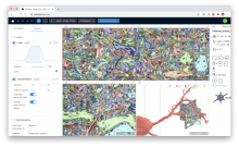
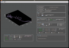
ClearMap is a toolbox for the analysis and registration of volumetric data from cleared tissues.

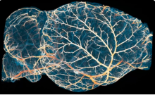
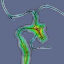
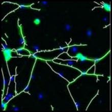
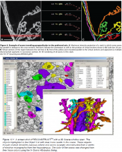
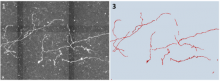
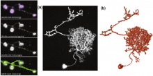
It was initially developed to map brain activity at cellular resolution in whole mouse brains using immediate early gene expression. It has since then been extended as a tool for the qunatification of whole mouse brain vascualtur networks at capilary resolution.

It is composed of sevral specialized modules or scripts: tubemap, cellmap, WobblyStitcher.

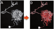
ClearMap has been designed to analyze O(TB) 3d datasets obtained via light sheet microscopy from iDISCO+ cleared tissue samples immunolabeled for proteins. The ClearMap tools may also be useful for data obtained with other types of microscopes, types of markers, clearing techniques, as well as other species, organs, or samples.

ClearMap SCreenshot
Description

Phindr3D is a comprehensive shallow-learning framework for automated quantitative phenotyping of three-dimensional (3D) high content screening image data using unsupervised data-driven voxel-based feature learning, which enables computationally facile classification, clustering and data visualization.

Please see our GitHub page and the original publication for details.

Description

webKnossos is an open-source data sharing and annotation platform for tera-scale 2D and 3D image datasets.

The core features of webKnossos are:

  • fast 3D data streaming
  • share links to specific locations in the data
  • uniquely fast skeleton annotation (flight mode) and
  • efficient volume annotation
  • mesh rendering
  • collaboration and sharing tools

webKnossos facilitates image analysis workflows on multi-terabyte datasets, including visualization of raw and multi-modal microscopy data, distributed training data generation and proof-reading of automatic segmentation.

As a scientific resource, webknossos.org serves as a database for published image datasets including their annotations.


Description

Vaa3d BJUT Fast Marching Spanning Tree algorithm dockerised workflow for BIAFLOWS

need a thumbnail
Description

3D Neuron Tracing with a Dockerized version of Vaa3D MOST Raytracer.

need a thumbnail
Description

3D Neuron Tracing using Dockerized version of Vaa3D Minimum Spanning Tree (MST).

need a thumbnail
Description

Rivuletpy dockerised workflow for BIAFLOWS.

has topic
need a thumbnail
Description

Vaa3d All-Path-Pruning 2.0 (APP2) dockerised workflow for BIAFLOWS.

need a thumbnail
Description

Paintera is a general visualization tool for 3D volumetric data and proof-reading in segmentation/reconstruction with a primary focus on neuron reconstruction from electron micrographs in connectomics. It features/supports:

  •  Views of orthogonal 2D cross-sections of the data at arbitrary angles and zoom levels
  •  Mipmaps for efficient display of arbitrarily large data at arbitrary scale levels
  •  Label data
    •  Painting
    •  Manual agglomeration
    •  3D visualization as polygon meshes
      •  Meshes for each mipmap level
      •  Mesh generation on-the-fly via marching cubes to incorporate painted labels and agglomerations in 3D visualization. Marching Cubes is parallelized over small blocks. Only relevant blocks are considered (huge speed-up for sparse label data).

Paintera is implemented in Java and makes extensive use of the UI framework JavaFX

Paintera screenshot

CEM

Description

Computer-assisted Evaluation of Myelin formation (CEM) is a collection designed to automate myelin quantification. It requires use input to choose the best threshold values. The myelin is calculated as an overlap between neuronal signal and oligodendrocyte signal. Results are given as pixel counts and percents.

CEM runs as an imageJ plugin with an optional Matlab extension to remove cell bodies. More details are published at Kerman et al. 2015 Development. Supplemental Material includes a detailed user manual and the download link.

Myelin
Description

vmtk is a collection of libraries and tools for 3D reconstruction, geometric analysis, mesh generation and surface data analysis for image-based modeling of blood vessels.

vmtk is composed of

  • C++ classes (VTK and ITK -based algorithms)
  • Python classes (high-level functionality - each class is a script)
  • PypeS - Python pipeable scripts, a framework which enables vmtk scripts to interact with each other


Description

"The plugin analyzes fluorescence microscopy images of neurites and nuclei of dissociated cultured neurons. Given user-defined thresholds, the plugin counts neuronal nuclei, and traces and measures neurite length."[...]" NeuriteTracer is a fast simple-to-use ImageJ plugin for the analysis of outgrowth in two-dimensional fluorescence microscopy images of neuronal cultures. The plugin performed well on images from three different types of neurons with distinct morphologies."

This plugin requires parameter setting: Threshold levels and scale (see more details on the related publication)

Description

We have developed a novel approach, named DF-Tracing, to tackle this challenge. This method first extracts the neurite signal (foreground) from a noisy image by using anisotropic filtering and automated thresholding. Then, DF-Tracing executes a coupled distance-field (DF) algorithm on the extracted foreground neurite signal and reconstructs the neuron morphology automatically. Two distance-transform based “force” fields are used: one for “pressure”, which is the distance transform field of foreground pixels (voxels) to the background, and another for “thrust”, which is the distance transform field of the foreground pixels to an automatically determined seed point. The coupling of these two force fields can“push” a “rolling ball” quickly along the skeleton of a neuron, reconstructing the 3D cell morphology.

Simple Tracing - DT-fields
Description

nctuTW is a "high-throughput computer method of reconstructing the neuronal structure of the fruit fly brain. The design philosophy of the proposed method differs from those of previous methods. We propose first to compute the 2D skeletons of a neuron in each slice of the image stack. The 3D neuronal structure is then constructed from the 2D skeletons. Biologists tend to use confocal microscopes for optimal images in a slice for human visualization; and images in two consecutive slices contain overlapped information. Consequently, a spherical object becomes oval in the image stack; that is, neurons in the image stack do not reflect the true shape of the neuron. This is the main reason we chose not to work directly on the 3D volume.

The proposed method comprises two steps. The first is the image processing step, which involves computing a set of voxels that is a superset of the 3D centerlines of the neuron. The shortest path graph algorithm then computes the centerlines. The proposed method was applied to process more than 16 000 neurons. By using a large amount of reconstructions, this study also demonstrated a result derived from the reconstructed data using the clustering technique." (Extracted from reference publication)

Illustrative image shows gold standard (top) and method results (bottom). 

nctuTW_results_example
Description

By combining multiple image alignment and tracing into one program, Reconstruct (TM) allows images to be processed more efficiently. Tracing can be done directly on the transformed images and alignments can be asily modified. Reconstruct (TM) was developed from years of experience working with high magnification serial section images of brain tissue. (Extracted from User Manual)

"The original platform of the Reconstruct program allows a user to trace objects in serial sections by manually drawing the outline of each object on each section, which is time-consuming. We modified Reconstruct to enable semi-automatic tracing of axons using a region-growing algorithm called wildfire."

Reconstruct_standaloneapp_example_Results
Description

JFilament is an ImageJ plugin for segmentation and tracking of 2D and 3D filaments in fluorescenece microscopy images. The main algorithm used in Jfilament is "Stretching Open Active Contours" (SOAC). In order to use this method, the user must define seed points in the image where the SOAC method will begin.

JFilament also includes 2D "closed" active contours which can be used for tasks such as segmentation and tracking of cell boundaries.


JFilament_ImageJ_pulgin_Window
Description

ORION: Online Reconstruction and functional Imaging Of Neurons: segmentation and tracing of neurons for reconstruction.

A project to develop tools that explore single neuron function via sophisticated image analysis. ORION software bridges advanced optical imaging and compartmental modeling of neuronal function by rapidly, accurately, and robustly generating, from structural image data, a cylindrical morphology model suitable for simulating neuronal function. The goal of this project is to develop a computational and experimental framework to allow real-time mapping of functional imaging data (e.g., spatio-temporal patterns of dendritic voltages or intracellularions) to neuronal structure, during the very limited duration of an acute experiment.

ORION_example_result
Description

The invention comprises a software tool, NeuronMetrics, which functions as a set of modules that run in the open-source program ImageJ. NeuronMetrics features a novel method for estimating neural “branch number” (a measure of the axonal complexity) from two-dimensional images. In addition, the tool features a novel method for modeling neural structure in large “gaps” that result from image artifacts.


has topic
need a thumbnail
Description

Neural Circuit Tracer (NCTracer) is open source software for automated and manual tracing of neurites from light microscopy stacks of images. NCTracer has more than one workflow available for neuron tracing. 


"The Neural Circuit Tracer is open source software built using Java (Sun Microsystems) and Matlab (MathWorks, Inc., Natick MA). It is based on the core of ImageJ (http://rsbweb.nih.gov/ij) and the graphic user interface has been developed by using Java Swings. The software combines anumber of functionalities of ImageJ with several newly developed functions for automated and manual tracing of neurites. The Neural Circuit Tracer is designed in a way
that will allow the users to add any plug-ins developed for ImageJ. More importantly, functions written in MatLab and converted into Java with Matlab JA toolbox can also be added to the Neural Circuit Tracer." 

Example of output from Neural Circuit Tracer
Description

hIPNAT (hIPNAT: Image Processing for NeuroAnatomy and Tree-like structures) is a set of tools for the analysis of images of neurons and other tree-like morphologies. It is written for ImageJ, the de facto standard in scientific image processing. It is available through the ImageJ Neuroanatomy update site.

need a thumbnail
Description

"we propose a novel automatic 3D neuron reconstruction algorithm, named Rivulet, which is based on the multi-stencils fast-marching and iterative back-tracking. The proposed Rivulet algorithm is capable of tracing discontinuous areas without being interrupted by densely distributed noises." 

This plugin can be used with default parameters or with user-defined parameters.

Example image obtained from Rivulet Wiki website (https://github.com/RivuletStudio/Rivulet-Neuron-Tracing-Toolbox/wiki

Traceplot_Rivulet
Description

"we present a new fully automated 3D reconstruction algorithm, called TReMAP, short for Tracing, Reverse Mapping and Assembling of 2D Projections. Instead of tracing a 3D image directly in the 3D space as seen in majority of the tracing methods, we first trace the 2D projection trees in 2Dplanes, followed by reverse-mapping the resulting 2D tracing results back into the 3D space as 3D curves; then we use a minimal spanning tree (MST) method to assemble all the 3D curves to generate the final 3D reconstruction. Because we simplify a 3D reconstruction problem into 2D, the computational costs are reduced dramatically." 

Suitable for high throughput neuron image analysis (image sizes >10GB). This plugin can be used with default parameters or user-defined parameters.

Example_TReMAP_Result
Description

All-path-pruning 2.0 (APP2) is a component of Vaa3D. APP2 prunes an initial reconstruction tree of a neuron’s morphology using a long-segment-first hierarchical procedure instead of the original termini-first-search process in APP. APP2 computes the distance transform of all image voxels directly for a gray-scale image, without the need to binarize the image before invoking the conventional distance transform. APP2 uses a fast-marching algorithm-based method to compute the initial reconstruction trees without pre-computing a large graph. This method allows to trace large images. This method can be used with default parameters or user-defined parameters.

APP2_Vaa3D_example_Result
Description

"We have developed an automatic graph algorithm, called the all-path pruning (APP), to trace the 3D structure of a neuron. To avoid potential mis-tracing of some parts of a neuron, an APP first produces an initial over-reconstruction, by tracing the optimal geodesic shortest path from the seed location to every possible destination voxel/pixel location in the image. Since the initial reconstruction contains all the possible paths and thus could contain redundant structural components (SC), we simplify the entire reconstruction without compromising its connectedness by pruning the redundant structural elements, using a new maximal- covering minimal-redundant (MCMR) subgraph algorithm. We show that MCMR has a linear computational complexity and will converge. We examined the performance of our method using challenging 3D neuronal image datasets of model organisms (e.g. fruit fly)"

This plugin can be used with default parameters or user-defined parameters.

APP_Vaa3D_example_results
Description

The Sprout Morphology plugin measures sprout number, length, width and cell density of endothelial cell (EC) sprouts grown in a bead sprouting assay. It optionally includes measuring the coverage of these sprouts with pericytes included in the assay, as well as the endothelial cell/pericyte ratio.

graphical abstract
Description

We propose to use a kernel density estimation (KDE) based approach for classification. This non-parametric approach intrinsically provides the likelihood of membership for each class in a principled manner. The implementation was used in Ghani2016. Any papers using this code should cite Ghani2016 accordingly. The software has been tested under Matlab R2013b.


Sample Data: Annotated two-photon images of dendritic spines

Description

Neurolucida is a powerful tool for creating and analyzing realistic, meaningful, and quantifiable neuron reconstructions from microscope images. Perform detailed morphometric analysis of neurons, such as quantifying 1) the number of dendrites, axons, nodes, synapses, and spines, 2) the length, width, and volume of dendrites and axons, 3) the area and volume of the soma, and 4) the complexity and extension of neurons. See 10.3389/fnins.2012.00049

Neurolucida example
Description

neuTube is a collection of neuron reconstruction tools from fluorescence microscope images. It has an interactive system with a 3D viewer, which can be clicked in 3D and perform neuron tracing automatically and semi-automatically. It can automatically recognize branching points as junctions. Traced neurons can be exported to swc format, which could be imported by various software packages. neuTube has Win and Mac OS standalone executable builds and may also be installed by manual compilation. In addition, neuTube can be used as a plugin in Vaa3D.


Neutube_standaloneapp_window_overview
Description

Neuron studio is a software package to reconstruct neurons from 3D confocal images. Reconstruction can be done manually, semi-manually or fully automatic. The images as well as the detected objects are rendered in 3D. A spine detection and classification function is also included. Results can be exported as a text file with coords of the spines. It seems that active development has stopped in 2009. NeuronStudio is being developed at the Computational Neurobiology and Imaging Center (CNIC), a research laboratory at the Neuroscience Department of the Mount Sinai School of Medicine in New York.

NeuronStudio can be used with default parameters or user-defined parameters (Fully or semi-automated).

NeuronStudio_standaloneapp_window_overview
Description

not available anymore in version 3.0 and up?

The forum discussion below might be of interest. Sample cppproj files are available in the discussion.

Measure Neurons - Total Neuron Length Measurement

need a thumbnail