Image registration

Image registration is the process of transforming different sets of data into one coordinate system. Registration is necessary in order to be able to compare or integrate the data obtained from different sensors/imaging modalities, at different times, from different view points, etc. . Registration can be based on correspondence established between the landmarks or feature points. Alternatively, some similarity/distance metric is established between the image intensity maps to navigate the registration process.

Synonyms
Image alignment
Description

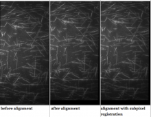
Fast4DReg is a Fiji macro for drift correction for 2D and 3D video and is able to correct drift in all x-, y- and/or z-directions. Fast4DReg creates intensity projections along both axes and estimates their drift using cross-correlation based drift correction, and then translates the video frame by frame. Additionally, Fast4DReg can be used for alignment multi-channel 2D or 3D images which is particularly useful for instruments that suffer from a misalignment of channels.

has function
Description


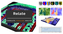
Relate is a correlative software package optimised to work with EM, EDS, EBSD, & AFM data and images.  It provides the tools you need to correlate data from different microscopes, visualise multi-layered data in 2D and 3D, and conduct correlative analyses.

  • Combining data from different imaging modalities (e.g. AFM, EDS & EBSD)

  • Interactive display of multi-layer correlated data

  • Analytical tools for metadata interrogation

  • Documented workflows and processes

Correlate

  • Import data from AZtec using the H5oina file format
  • Import AFM data
  • Correlate both sets of data using intuitive image overlays and image matching tools
  • Produce combined multimodal datasets

Visualise

  • 2D display of multi-layered data
  • 3D visualisation of topography combined with AFM material properties, EM images, and EDS & EBSD map overlays
  • Customisation of colour palettes, data overlays, image rendering options, and document display
  • Export images and animations

Analyse

  • Generate profile (cross section) views of multimodal data
  • Measure and quantify data across multiple layers
  • Analyse areas via data thresholding using amount of x-ray counts, phase maps, height, or other material properties.
  • Select an extensive range of measurement parameters
  • Export analytical data to text or CSV files
Relate analysis workflow example
Description

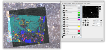
Correlia is an open-source ImageJ/FIJI plug-in for the registration of 2D multi-modal microscopy data-sets. The software is developed at ProVIS - Centre for Correlative Microscopy and is specifically designed for the needs of chemical microscopy involving various micrographs as well as chemical maps at different resolutions and field-of-views.

Correlia

MIA

Description

ModularImageAnalysis (MIA) is an ImageJ plugin which provides a modular framework for assembling image and object analysis workflows. Detected objects can be transformed, filtered, measured and related. Analysis workflows are batch-enabled by default, allowing easy processing of high-content datasets.

MIA is designed for “out-of-the-box” compatibility with spatially-calibrated 5D images, yielding measurements in both pixel and physical units.  Functionality can be extended both internally, via integration with SciJava’s scripting interface, and externally, with Java modules that extend the MIA framework. Both have full access to all objects and images in the analysis workspace.

Workflows are, by default, compatible with batch processing multiple files within a single folder. Thanks to Bio-Formats, MIA has native support for multi-series image formats such as Leica .lif and Nikon .nd2.

Workflows can be automated from initial image loading through processing, object detection, measurement extraction, visualisation, and data exporting. MIA includes near 200 modules integrated with key ImageJ plugins such as Bio-Formats, TrackMate and Weka Trainable Segmentation.

Module(s) can be turned on/off dynamically in response to factors such as availability of images and objects, user inputs and measurement-based filters. Switches can also be added to “processing view” for easy workflow control.

MIA is developed in the Wolfson Bioimaging Facility at the University of Bristol.

Description

ClearMap is a toolbox for the analysis and registration of volumetric data from cleared tissues.

It was initially developed to map brain activity at cellular resolution in whole mouse brains using immediate early gene expression. It has since then been extended as a tool for the qunatification of whole mouse brain vascualtur networks at capilary resolution.

It is composed of sevral specialized modules or scripts: tubemap, cellmap, WobblyStitcher.

ClearMap has been designed to analyze O(TB) 3d datasets obtained via light sheet microscopy from iDISCO+ cleared tissue samples immunolabeled for proteins. The ClearMap tools may also be useful for data obtained with other types of microscopes, types of markers, clearing techniques, as well as other species, organs, or samples.

ClearMap SCreenshot
Description

This Fiji plugin is a python script for CLEM registration using deep learning, but it could be applied in principle to other modalities. The pretrained model was learned on chromatin SEM images and fluorescent staining, but a script is also provided to train an new model, based on CSBDeep. The registration is then performed as a feature based registration, using register virtual stack plugin (which extract features and then perform RANSAc. Editing the script in python gives access to more option (such as the transformation model to be used, similarity by default. Images need to be prepared such that they contain only one channel, but channel of ineterst (to be transformed with the same transformation) can be given as input, and Transform Virtual Stack plugin can be used as well.

F1000R Figure 1 DeepCLEM
Description

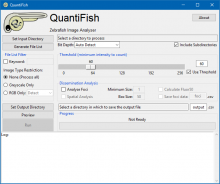
QuantiFish is a quantification program intended for measuring fluorescence in images of zebrafish, although use with images of other specimens is possible. This package is geared towards analysis of fluorescent infection models. The software is designed to automate processing of images of single fish, and outputs results as a .csv file. Alongside measures of total fluorescence above a threshold, this package also introduces several measures for dissemination and distribution of fluorescence throughout the specimen.

QuantiFish User Interface
Description

Epina ImageLab is a Microsoft Windows-based multisensor imaging tool for processing and analyzing hyperspectral images. It is a modular system consisting of a basic engine, a graphical user interface, a chemometrics toolbox and optional user-supplied modules. It supports the most important spectroscopic imaging techniques, such as UV/Vis, infrared, Raman, THz, optical emission/absorption, and mass spectrometry. On top of that Epina ImageLab enables the user to merge hyperspectral images with maps of physical properties and conventional high-resolution color photos. 

need a thumbnail
Description

Align two images using intensity correlation, feature matching, or control point mapping

Together, Image Processing Toolbox™ and Computer Vision Toolbox™ offer four image registration solutions: interactive registration with a Registration Estimator app, intensity-based automatic image registration, control point registration, and automated feature matching. 

has topic
need a thumbnail
Description

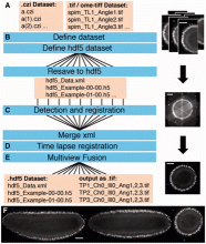
Automated workflow for performing multiview reconstruction of large multiview, multichannel, multiillumination time-lapse SPIM data on a high performance computing (HPC) cluster or on a single workstation. 

Description

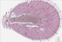
This python toolbox performs registration between 2-D microscopy images from the same tissue section or serial sections in several ways to achieve imaging mass spectrometry (IMS) experimental goals.

This code supports the following works and enables others to perform the workflows outlined in the following works, please cite them if you use this toolbox:

  • Advanced Registration and Analysis of MALDI Imaging Mass Spectrometry Measurements through Autofluorescence Microscopy10.1021/acs.analchem.8b02884

  • Next Generation Histology-directed Imaging Mass Spectrometry Driven by Autofluorescence Microscopy10.1021/acs.analchem.8b02885

need a thumbnail
Description

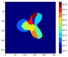
The Binary Pattern Dictionary Learning (BPDL) package is suitable for image analysis on a set/sequence of images to determine an atlas of a compact region. In particular, the application can be maping gene activation accross many samples, brain activations in a time domain, etc.

Atlas
Description

BIRL stands for "Benchmark on Image Registration methods with Landmark validation". BIRL is a cross-platform framework for comparison of image registration methods with landmark validation (registration precision is measured by user landmarks). The project contains a set of sample images with related landmark annotations and experimental evaluation of state-of-the-art image registration methods.

Some key features of the framework:

  • automatic execution of image registration of a sequence of image pairs
  • integrated evaluation of registration performances using Target Registration Error (TRE)
  • integrated visualization of performed registration
  • running several image registration experiment in parallel
  • resuming unfinished sequence of registration benchmark
  • handling around dataset and creating own experiments
  • rerun evaluation and visualisation for finished experiments
has topic
has function
Description

It is an interactive front-end visualization for registration software based on Elasix (VTK/ITK)

has topic
need a thumbnail
Description

Non linear registration intensity based for MRI brain exams. To be applied after FLIRT

a brain mri
Description

FLIRT (FMRIB's Linear Image Registration Tool) is a fully automated robust and accurate tool for linear (affine) intra- and inter-modal brain image registration.

FLIRT comes with a main GUI as well as three supporting guis:

  • ApplyXFM - for applying saved transformations and changing FOVs
  • InvertXFM - for inverting saved transformations
  • ConcatXFM - for concatenating saved transformations
Description

SimpleITK provides a simplified interface to ITK in a variety of languages. A user can either download pre-built binaries, if they are available for the desired platform and language, or SimpleITK can be built from the source code. Currently, Python binaries are available on Microsoft Windows, GNU Linux and Mac OS X. C# and Java binaries are available for Windows. We are also working towards supporting R packaging.

need a thumbnail
Description

Python/C++ port of the ImageJ extension TurboReg/StackReg written by Philippe Thevenaz/EPFL.

A python extension for the automatic alignment of a source image or a stack (movie) to a target image/reference frame.

need a thumbnail
Description

Align_slices in stack utilized the template matching function cvMatch_Template to do slice registration(alignment) based on a selected landmark.
This function will try to find the landmark or the most similar image pattern in every slice and translate each slice so that the landmark pattern will be the same position throughout the whole stack. It could be used to fix the drift of a time-lapse image stacks.

Source code: link

Input data: image stack
output data: image stack

has function
Description

This ImageJ plugin contains two functions. The first one is the cvMatch_Template. It implements the template matching function from the OpenCV library. The second function Align_slices in stack utilized the previous matching function to do slice registration(alignment) based on a selected landmark. 

For more details, refer to the page of each component. 

cvMatch_Template

Align Slices in Stack

has function
Description

Elastix is a toolbox for rigid and nonrigid registration of (medical) images.

Elastix is based on the ITK library, and provides additional algorithms for image registration. 

The software can be run as a single-line command, making it easy to include in larger scripts or workflows. The user needs to edit a configuration file that contains all relevant parameters for registration: transformation model, metric used to comapre images, optimization algorithm, mutliscale pyramidal representation of images...

Nowadays elastix is accompanied by SimpleElastix, making it available in other languages like C++, Python, Java, R, Ruby, C# and Lua.

elastix logo
Description

CaPTk is a software platform for analysis of radiographic cancer images, currently focusing on brain, breast, and lung cancer. CaPTk integrates advanced, validated tools performing various aspects of medical image analysis, that have been developed in the context of active clinical research studies and collaborations toward addressing real clinical needs. With emphasis given in its use as a very lightweight and efficient viewer, and with no prerequisites for substantial computational background, CaPTk aims to facilitate the swift translation of advanced computational algorithms into routine clinical quantification, analysis, decision making, and reporting workflow. Its long-term goal is providing widely used technology that leverages the value of advanced imaging analytics in cancer prediction, diagnosis and prognosis, as well as in better understanding the biological mechanisms of cancer development.

CaPTk
Description

Reconstruct big images from overlapping tiled images on a Spark cluster.

The code is based on the Stitching plugin for Fiji https://github.com/fiji/Stitching

SparkStitching