Machine learning

Synonyms
Artificial intelligence
Description

BIIGLE is a web-based software for image and video annotation that enables collaborative research on large datasets. It offers tools for manual and computer-assisted annotation, quality control and the collaboration on custom taxonomies to describe objects. BIIGLE is freely available and can be installed in cloud environments, a local network or on mobile platforms during research expeditions. The public instance on biigle.de is free for non-commercial use.

BIIGLE Logo

Metric Reloaded: how to select and use your metrics

Submitted by Perrine on Wed, 02/14/2024 - 13:39

The mission of Metrics Reloaded is to guide researchers in the selection of appropriate performance metrics for biomedical image analysis problems, as well as provide a comprehensive online resource for metric-related information and pitfalls. This website provides ressources such as a tool to select the best metric, as well as tutorials about the way to use and interpret metrics in image analysis.


Description

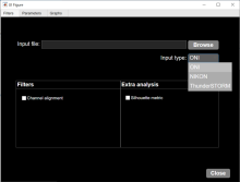
MATLAB app to characterize nanoparticles imaged with super-resolution microscopy. nanoFeatures will read text and csv files from the NIKON and ONI microscopes and from the ThunderSTORM Fiji plugin, then cluster the localizations and filter by size and sphericity and finally output nanoparticle features like size, aspect ratio, and number of localizations per cluster (total and for each channel).

GUI first tab to browse and input files, select input type and check extra filters if needed.
Description

ELEPHANT is a platform for 3D cell tracking, based on incremental and interactive deep learning.
It implements a client-server architecture. The server is built as a web application that serves deep learning-based algorithms. The client application is implemented by extending Mastodon, providing a user interface for annotation, proofreading and visualization.

from https://elephant-track.github.io/#/v0.5/?id=_5-proofreading
Description

ZeroCostDL4Mic: exploiting Google Colab to develop a free and open-source toolbox for Deep-Learning in microscopy

ZeroCostDL4Mic is a collection of self-explanatory Jupyter Notebooks for Google Colab that features an easy-to-use graphical user interface. They are meant to quickly get you started on learning to use deep-learning for microscopy. 

need a thumbnail
Description

btrack is a Python library for multi object tracking, used to reconstruct trajectories in crowded fields. btrack implemented a residual U-Net model coupledd with a classification CNN to allow accurate instance segmentation of the cell nuclei. To track the cells over time and through cell divisions, btrack developed a Bayesian cell tracking methodology that uses input features from the images to enable the retrieval of multi-generational lineage information from a corpus of thousands of hours of live-cell imaging data.

need a thumbnail
Description

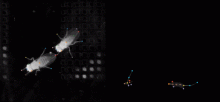
Open source deep learning based framework for multi-animal pose tracking. It can track animal and any number of animals and has a labeling/training GUI for learning and proofreading.

has topic
has function
Description

Algorithm and software created to extract animal trajectories from videos of a collection of animals up to 100 individuals. Idtrackerai uses two convolutional networks: one for animal identification and another to detect when animals touch or cross each other.

has topic
has function
Description

The method proposed in this paper is a robust combination of multi-task learning and unsupervised domain adaptation for segmenting amoeboid cells in microscopy. This end-to-end framework provides a consolidated mechanism to harness the potential of multi-task learning to isolate and segment clustered cells from low contrast brightfield images, and it simultaneously leverages deep domain adaptation to segment fluorescent cells without explicit pixel-level re- annotation of the data.

The entry-point to the codebase is the main.py file. The user has the option to

  • Train the network on their own dataset
  • Load a pre-trained model and use that for inference on their own data
  • NoteThe provided pretrained model was trained on 256x256 images. Results on different resolutions could require fine-tuning This model is trained (supervised) on brightfield, and domain adapted to fluorescence data. The results are saved as 'inference.png'
has function
daman
Description

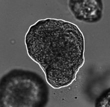
This workflow describes a deep-learning based pipeline for reliable single-organoid segmentation and tracking in 2D+t high-resolution brightfield microscopy of mouse mammary epithelial organoids. The pipeline involves a four-layer U-Net to infer semantic segmentation predictions, adaptive morphological filtering to establish candidate organoid instances, and a shape-similarity-constrained, instance-segmentation-correcting tracking step to associate the corresponding organoid instances in time.

It is particularly focused on automatically detecting an organoid located approximately in the center of the first frame and track all its subsequent instances in the remaining frames, emphasizing on accurate organoid boundary delineation. Furthermore, segmentation network was trained using plausible pix2pixHD-generated bioimage data. Syntheric image simulator code and data are also available here.

Adapted from https://cbia.fi.muni.cz/research/spatiotemporal/organoids.html
Description

OrganoID is an image analysis platform that automatically recognizes, labels, and tracks single organoids, pixel-by-pixel, in brightfield and phase-contrast microscopy experiments. The platform was trained on images of pancreatic cancer organoids and validated on separate images of pancreatic, lung, colon, and adenoid cystic carcinoma organoids.

need a thumbnail
Description

Fractal is a framework to process high-content imaging data at scale and prepare it for interactive visualization. Fractal provides distributed workflows that convert TBs of image data into OME-Zarr files. The platform then processes the 3D image data by applying tasks like illumination correction, maximum intensity projection, 3D segmentation using cellpose and measurements using napari workflows. The pyramidal OME-Zarr files enable interactive visualization in the napari viewer.

need a thumbnail
Description

This workflow applies a Stardist pre-trained model (versatile_fluo or versatile_HE) depending on the input images ie. uses both models for a dataset including both fluorescence (grayscale or RGB where all channels are equal) and H&E stained (RGB where channels are not equal) images.

This version uses tensorflow CPU version (See Dockerfile) to ensure compatibility with a larger number of computers. A GPU version should be possible by adapting the Dockerfile with tensorflow-gpu and/or nvidia-docker images.

has topic
has function
need a thumbnail
Description

This workflow processes a group of images containing cells with discernible nuclei and segments the nuclei and outputs a binary mask that show where nuclei were detected. It performs 2D nuclei segmentation using pre-trained nuclei segmentation models of Cellpose. And it was developed as a test workflow for Neubias BIAFLOWS Benchmarking tool.

has topic
has function
need a thumbnail
Description

The Incucyte® Base Analysis Software provides a guided interface and purpose-built tools, which include the process of acquiring, viewing, analyzing and sharing images of living cells.

need a thumbnail
Description

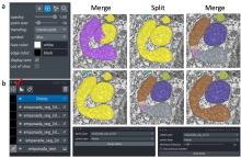
The empanada-napari plugin is built to democratize deep learning image segmentation for researchers in electron microscopy (EM). It ships with MitoNet, a generalist model for the instance segmentation of mitochondria. There are also tools to quickly build and annotate training datasets, train generic panoptic segmentation models, finetune existing models, and scalably run inference on 2D or 3D data. To make segmentation model training faster and more robust, CEM pre-trained weights are used by default. These weights were trained using an unsupervised learning algorithm on over 1.5 million EM images from hundreds of unique EM datasets making them remarkably general.

Empanada-napari
Description

AnnotatorJ is a Fiji Plugin to ease annotation of images, particulrly useful for Deep Learning or to validate an alogorithm. Interestingly, it allows annotation for instance segmentation, semantic segmentation, or bounding box annotations. It includes toolssuch as active contours to ease these annotations.

has topic
has function
annotatorJ
Description

Machine Learning made easy

APEER ML provides an easy way to train your own machine learning
models and segment your microscopy images. No expertise or coding required.

APEER

Description

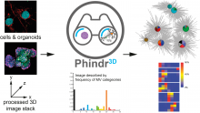
Phindr3D is a comprehensive shallow-learning framework for automated quantitative phenotyping of three-dimensional (3D) high content screening image data using unsupervised data-driven voxel-based feature learning, which enables computationally facile classification, clustering and data visualization.

Please see our GitHub page and the original publication for details.

Data Science in Cell Imaging (DSCI) course material

Submitted by assafzar on Thu, 12/31/2020 - 10:08

Graduate course at the department of Software and Information Systems Engineering at Ben-Gurion University of the Negev.

The recent explosion in high-content, dynamic and multidimensional imaging data is transforming cell imaging into a “Data Science” field. This course will review the state-of-the-art in visualizing, processing, integrating and mining massive cell image data sets, deciphering complex patterns and turning them into new biological insight. It will include a mix of approaches in computer science, machine learning and computer vision applied to bio-imaging data.

Description

Deep learning based image restoration methods have recently been made available to restore images from under-exposed imaging conditions, increase spatio-temporal resolution (CARE) or self-supervised image denoising (Noise2Void). These powerful methods outperform conventional state-of-the-art methods and leverage down-stream analyses significantly such as segmentation and quantification.

To bring these new tools to a broader platform in the image analysis community, we developed a simple Jupyter based graphical user interface for CARE and Noise2Void, which lowers the burden for non-programmers and biologists to access these powerful methods in their daily routine.  CARE-less supports temporal, multi-channel image and volumetric data and many file formats by using the bioformats library. The user is guided through the different computation steps via inline documentation. For standard use cases, the graphical user interface exposes the most relevant parameters such as patch size and number of training iterations, while expert users still have access to advanced parameters such as U-net depth and kernel sizes. In addition, CARE-less provides visual outputs for training convergence and restoration quality. Any project settings can be stored and reused from command line for processing on compute clusters. The generated output files preserve important meta-data such as pixel sizes, axial spacing and time intervals.

need a thumbnail
Description

This tool allows the user to define structures of interest by interactively marking a subset of pixels. Thanks to the real-time feedback, the user can place new markings strategically, depending on the current outcome.

Description

BioImage.IO -- a collaborative effort to bring AI models to the bioimaging community. 

  • Integrated with Fiji, ilastik, ImJoy and more
  • Try model instantly with BioEngine
  • Contribute your models via Github

This is a database of pretrained deep Learning models. 

need a thumbnail
Description

Yet another pixel classifier Yapic is a deep learning tool to :

train your own filter to enhance the structure of your choice 

train multiple filter at once 

it is based on the u-net convolutional filter . 

To train it : annotation can come from example from Ilastik software , tif labelled files can be transferred to yapic. 

Training takes about hours to days , prediction takes seconds once trained .

It can be ran from command line .

note that only 10 to 20 images with sparse labeling are required for efficient training 

has function
need a thumbnail
Description

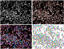
LOBSTER (Little Objects Segmentation and Tracking Environment), an environment designed to help scientists design and customize image analysis workflows to accurately characterize biological objects from a broad range of fluorescence microscopy images, including large images, i.e. terabytes of data, exceeding workstation main memory.

  • 75 workflows available 
  • no programming, with GUI
  • matlab based 
Description

Web based application for deep learning cell classifier . Aim to replace cellprofiler analyst and advanced cell classifier.

has topic
has function
need a thumbnail
Description

This is the ImageJ/Fiji plugin for StarDist, a cell/nuclei detection method for microscopy images with star-convex shape priors ( typically for Dapi like staining of nuclei). The plugin can be used to apply already trained models to new images.

Stardist
Description

Summary

Deep learning-based segmentation of cells, both fluorescence, and bright-field images ("a generalist algorithm for cellular segmentation"). The tool can be used either online or local or via notebooks (e.g. ZeroCostDL4Mic).

How to use it

cellpose can be used online via ready-to-use Jyupyter notebooks with very good documentation. These notebooks are listed here.

Local Installation

The general local installation procedure can be found here.

... Installing to Silicon Mac (M1 processor) needs some tricks, and as of October 2021, the following sequence of commands works. numba should be conda-installed before pip-installing the cellpose.


conda create --name cellpose python=3.8
conda activate cellpose
conda install numba
git clone https://github.com/MouseLand/cellpose.git
cd cellpose
pip install -e .

has topic
has function
Description

DeepImageJ is a user-friendly plugin that enables the use of a variety of pre-trained deep learning models in ImageJ and Fiji. The plugin bridges the gap between deep learning and standard life-science applications. DeepImageJ runs image-to-image operations on a standard CPU-based computer and does not require any deep learning expertise.

Training developper constructs and upload trained model, and made them available to users.

Models are available in a repository here.

It is macro recordable. It is advised to use DeepImageJ on a computer with GPU (CPU will likely be 20x slower)

has topic
deepImageJ
Description

Rivuletpy dockerised workflow for BIAFLOWS.

has topic
need a thumbnail
Description

Cell tracking using MU-Lux-CZ algorithm. Dockerized Workflow for BIAFLOWS implemented by Martin Maska (Masaryk University).

has topic
has function
need a thumbnail
Description

Track non-dividing particles in 2D time-lapse image.

has topic
has function
need a thumbnail
Description

Execute Nuclei Segmentation in 3D images using pixel classification with ilastik.

has topic
has function
need a thumbnail
Description

U-Net segmentation as presented in Reference Publication. The model predicts three classes: background, edge and foreground. The model was trained with Kaggle Data Science Bowl (DSB) 2018 training set.

has topic
has function
need a thumbnail
Description

Nuclei Segmentation using Deep Learning for individual cell analysis (DeepCell).

has topic
has function
need a thumbnail
Description

Wavelet Toolbox™ provides apps and functions for the time-frequency analysis of signals and multiscale analysis of images.

need a thumbnail
Description

ASAP allows to automatically detect, classify and quantify structures acquired by super resolution microscopy. 

Description

This plugin ships automated methods for extracting trajectories of multiples objects in a sequence of 2D or 3D images. Up to version 2 it was known as the ‘Probabilistic particle tracker’ plugin.

need a thumbnail
Description

Wolfram Mathematica (usually termed Mathematica) is a modern technical computing system spanning most areas of technical computing — including neural networksmachine learningimage processinggeometrydata sciencevisualizations, and others. The system is used in many technical, scientific, engineering, mathematical, and computing fields.

Description

Dragonfly is a software platform for the intuitive inspection of multi-scale multi-modality image data. Its user-friendly experience translates into powerful quantitative findings with high-impact visuals, driven by nuanced easy-to-learn controls.

For segmentation: It provides an engine fior machine Learning, Watershed and superpixel methods, support histological data .

It offers a 3D viewer, and python scripting capacities .

It is free for reserach use, but not for commercial usage.

DragonFly
Description

online image data management system which supports authenticated image upload, cloud-based storage, project-based management and viewing of standard and whole slide images. One can use different annotation tools to highlight important objects or areas within images. It is the first basic version and new features such as sharing for easy collaboration with your colleagues or first automated analysis applications based on artificial intelligence will be added soon.

Ikosa Portal: multi user image data management

Ikosa Prisma: Automated Image Analysis based on deep learning (available in summer 2019)

Free if limited to 2 users and 1 gigabyte, otherwise montly fees.


ikosa
Description

NEUBIAS-WG5 workflow for nuclei segmentation using ilastik v1.3.2 and Python post-processing.

has topic
has function
need a thumbnail
Description

NEUBIAS-WG5 workflow for nuclei segmentation using Mask-RCNN. The workflow uses Matterport Mask-RCNN. Keras implementation. The model was trained with Kaggle 2018 Data Science Bowl images.

has topic
need a thumbnail
Description

This workflow predict landmark positions on images by using DMBL landmark detection models.

has topic
has function
need a thumbnail