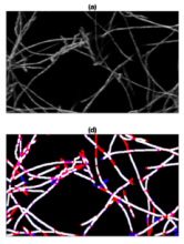
Component

A Component is an implementation of certain image processing / analysis algorithms.

Each component alone does not solve a Bioimage Analysis problem.

These problems can be addressed by combining such components into workflows.

Description

HistoMetrix is an advanced histology analysis software designed to simplify image processing and analysis for biologists and pathologists. Powered by the most advanced deep learning technology, HistoMetrix enables you to effortlessly uncover valuable insights and visualize results without the need for extensive technical expertise. Let’s explore the key features that make HistoMetrix the ultimate solution for histology analysis.

HistoMetrix leverages cutting-edge deep learning technology to streamline and simplify image processing for histology analysis. You can easily navigate through complex datasets, detect and analyze tissue structures, and extract meaningful information with just a few clicks. Research use only.


HistoMetrix combines advanced deep learning technology with cost-effectiveness, making it the ideal histology analysis software for biologists and pathologists. With HistoMetrix, effortlessly uncover valuable insights, visualize results, and simplify image processing, all while enjoying significant cost savings compared to other solutions. Benefit from affordable pricing plans, no expensive hardware requirements, and no need for costly training programs. HistoMetrix streamlines workflows, automates tasks, and provides efficient analysis tools, allowing you to save valuable time and resources. Experience the cost-effective solution for histology analysis and accelerate your research with HistoMetrix.

HistoMetriX for image analysis of histology slides

Metric Reloaded: how to select and use your metrics

Submitted by Perrine on Wed, 02/14/2024 - 13:39

The mission of Metrics Reloaded is to guide researchers in the selection of appropriate performance metrics for biomedical image analysis problems, as well as provide a comprehensive online resource for metric-related information and pitfalls. This website provides ressources such as a tool to select the best metric, as well as tutorials about the way to use and interpret metrics in image analysis.

Description

CellStich proposes a set of tools for 3D segmentation from 2D segmentation: it reassembles 2D labels obtained from cell in slices in unique 3D labels across slices. It isparticularly robust to anisotropy, and is the ideal companion to cellpose 2D models or other 2D deep learning based models. One could also think about using it for cell tracking by overlap (using time as a third dimension).

cellstitch
Description

Image segmentation and object detection performance measures

The goal of this package is to provide easy-to-use tools for evaluation of the performance of segmentation methods in biomedical image analysis and beyond, and to fasciliate the comparison of different methods by providing standardized implementations. This package currently only supports 2-D image data.

has function
Description

MATLAB app to characterize nanoparticles imaged with super-resolution microscopy. nanoFeatures will read text and csv files from the NIKON and ONI microscopes and from the ThunderSTORM Fiji plugin, then cluster the localizations and filter by size and sphericity and finally output nanoparticle features like size, aspect ratio, and number of localizations per cluster (total and for each channel).

GUI first tab to browse and input files, select input type and check extra filters if needed.