Clustering

Description

MATLAB app to characterize nanoparticles imaged with super-resolution microscopy. nanoFeatures will read text and csv files from the NIKON and ONI microscopes and from the ThunderSTORM Fiji plugin, then cluster the localizations and filter by size and sphericity and finally output nanoparticle features like size, aspect ratio, and number of localizations per cluster (total and for each channel).

GUI first tab to browse and input files, select input type and check extra filters if needed.
Description

Phindr3D is a comprehensive shallow-learning framework for automated quantitative phenotyping of three-dimensional (3D) high content screening image data using unsupervised data-driven voxel-based feature learning, which enables computationally facile classification, clustering and data visualization.

Please see our GitHub page and the original publication for details.

Description

Histology Topography Cytometry Analysis Toolbox (histoCAT) is a package to visualize and analyse multiplexed image cytometry data interactively. It can also export data in.fcs data for further analysis using  a specialized cytometry sofwtare such as Flowjo. 

It can be run as a compiled standalone or from matlab.

Description

Analyze the clustering behavior of nuclei in 3D images. The centers of the nuclei are detected. The nuclei are filtered by the presence of a signal in a different channel. The clustering is done with the density based algorithm DBSCAN. The nearest neighbor distances between all nuclei and those outside and inside of the clusters are calculated.

has function
Description

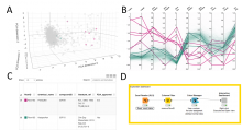
KNIME workflow to visualize a dataset described by multiple quantitative features (ex: a list of samples or cells, each described with multiple morphological features) as a 3D cloud of points (each point corresponding to one sample/cell) as well as a line plot (1 line per sample/cell).

For the 3D plot, the workflow uses Principal Component Analysis (PCA) for dimensionality reduction, ie it simplifies the information for each sample from n-features to 3 pseudo-features which are used as x,y,z-coordinates for each sample. The original features should cover similar value range, to make sure the PCA is not biased towards the large values features. One option is to normalize the values (min/max or Z-score). 

Also make sure that the resulting PCA represents a decent % of the original data variance (at least 70%). Otherwise the PCA plot will not be representative of the original data-distribution. The % is shown in the title of the PCA plot.

The workflow is interactive and so selecting in one panel of the figure will highlight in the other panel too.

It was originally published for the visualization of phenotypic kidney features in zebrafish, but the workflow is generic by design and can be reused for any quantitative feature set. 

KNIME-Workflow