Image deconvolution

Description

FluoGAN is a fluorescence image deconvolution software combining the knowledge of acquisition physical model with gan. It takes a fluctuating sequence of blurred, undersampled and noisy images of the sample of interest  fixed sample as input from wide field or confocal and returns a super resolved image.

FluoGan
Description


Relate is a correlative software package optimised to work with EM, EDS, EBSD, & AFM data and images.  It provides the tools you need to correlate data from different microscopes, visualise multi-layered data in 2D and 3D, and conduct correlative analyses.

  • Combining data from different imaging modalities (e.g. AFM, EDS & EBSD)

  • Interactive display of multi-layer correlated data

  • Analytical tools for metadata interrogation

  • Documented workflows and processes

Correlate

  • Import data from AZtec using the H5oina file format
  • Import AFM data
  • Correlate both sets of data using intuitive image overlays and image matching tools
  • Produce combined multimodal datasets

Visualise

  • 2D display of multi-layered data
  • 3D visualisation of topography combined with AFM material properties, EM images, and EDS & EBSD map overlays
  • Customisation of colour palettes, data overlays, image rendering options, and document display
  • Export images and animations

Analyse

  • Generate profile (cross section) views of multimodal data
  • Measure and quantify data across multiple layers
  • Analyse areas via data thresholding using amount of x-ray counts, phase maps, height, or other material properties.
  • Select an extensive range of measurement parameters
  • Export analytical data to text or CSV files
Relate analysis workflow example
Description

Deep learning based image restoration methods have recently been made available to restore images from under-exposed imaging conditions, increase spatio-temporal resolution (CARE) or self-supervised image denoising (Noise2Void). These powerful methods outperform conventional state-of-the-art methods and leverage down-stream analyses significantly such as segmentation and quantification.

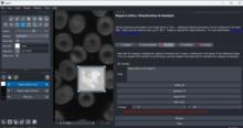
To bring these new tools to a broader platform in the image analysis community, we developed a simple Jupyter based graphical user interface for CARE and Noise2Void, which lowers the burden for non-programmers and biologists to access these powerful methods in their daily routine.  CARE-less supports temporal, multi-channel image and volumetric data and many file formats by using the bioformats library. The user is guided through the different computation steps via inline documentation. For standard use cases, the graphical user interface exposes the most relevant parameters such as patch size and number of training iterations, while expert users still have access to advanced parameters such as U-net depth and kernel sizes. In addition, CARE-less provides visual outputs for training convergence and restoration quality. Any project settings can be stored and reused from command line for processing on compute clusters. The generated output files preserve important meta-data such as pixel sizes, axial spacing and time intervals.

need a thumbnail
Description

Automated workflow for performing multiview reconstruction of large multiview, multichannel, multiillumination time-lapse SPIM data on a high performance computing (HPC) cluster or on a single workstation. 

Description

CLIJ2 is a GPU-accelerated image processing library for ImageJ/FijiIcy, Matlab and Java. It comes with hundreds of operations for filteringbinarizinglabelingmeasuring in images, projectionstransformations and mathematical operations for images. While most of these are classical image processing operations, CLIJ2 also allows performing operations on matrices potentially representing neighborhood relationships between cells and pixels.

CLIJ2 was developed to process images from fluorescence microscopy data of developing cells, tissues, organoids and organisms.