plugin

Description

This workflow predict landmark positions on images by using DMBL landmark detection models.

has topic
has function
need a thumbnail
Description

This workflow trains DMBL landmark detection models from a dataset of annotated images.

has function
need a thumbnail
Description

This workflow predict landmark positions on images by using LC landmark detection models.

has topic
has function
need a thumbnail
Description

This workflow trains LC landmark detection models from a dataset of annotated images.

has topic
has function
need a thumbnail
Description

This workflow predict landmark positions on images by using MSET landmark detection models.

has topic
has function
need a thumbnail
Description

This workflow trains MSET landmark detection models from a dataset of annotated images.

has topic
has function
need a thumbnail
Description

This workflow segments glands from H&E stained histopathological images
from the Gland Segmentation Challenge (GlaS2015) using deep learning (UNet).
UNet implementation largely inspired from PyTorch-UNet by Milesial. 

need a thumbnail
Description

ImageJ/FIJI plugin generating contour lines with equal spacing on top of an image (using overlay).

Description

jSLIC superpixels - is a segmentation method for clustering similar regions - superpixels - in the given image which are usually used for other segmentation techniques. The only two parameters are average (initial) size of each superpixel and rigidity parameter in range (0,1)

has topic
has function
superpixels - ROI
Description

This workflow processes a group of images containing cells with discernible nuclei and segments the nuclei and outputs a binary mask that show where nuclei were detected. It was developed as a test workflow for Neubias BIAFLOWS Benchmarking tool.

has function
Description

Runs fill holes operation on 3D images.

need a thumbnail
Description

This component convolves the image with maximum filter. Each voxel is set to the maximum value of its neighborhood. The neighborhood is defined by a kernel, which has a diameter of 3 voxels.

need a thumbnail
Description

ROI measurement plug-in for Icy.

has function
need a thumbnail
Description

This component convolves the image with minimum filter. Each voxel is set to the minimum value of its neighborhood. The neighborhood is defined by a kernel, which has a diameter of 3 voxels.

has function
need a thumbnail
Description

This workflow detects spots from a 3D image by using straightforward set of ImageJ components. It receives the Laplacian Radius and the Threshold  value s input.

has function
need a thumbnail
Description

This workflow detects spots in a 2D image by filtering the image by Laplacian of Gaussian (user defined radius) and detecting regional intensity minima (user defined noise tolerance).

has function
need a thumbnail
Description

Preprocessing step for high-density analysis methods in super resolution localisation microscopy: it aims at correcting artefacts due to these approaches with based on Haar Wavelet Kernel Analysis.

Description

The macro will segment nuclei and separate clustered nuclei in a 3D image using a distance transform watershed. As a result an index-mask image is written for each input image.

need a thumbnail
Description

This suite provides plugins to enhance 3D capabilities of ImageJ.

  • 3D Filters (mean, median, max, min, tophat, max local, …) and edge and symmetry filter
  • 3D Segmentation (iterative thresholding, spots segmentation, watershed, …)
    • 3D hysteresis thresholding with two thresholds (see 2D hysteresis for explanation).
    • 3D simple segmentation with thresholding to label 3D objects (similar to 3D objects counter).
    • 3D iterative thresholding (find optimal threshold for each object).
    • 3D spot segmentation with various local threshold estimations.
    • 3D Maxima Finder (with noise parameter)
    • 3D seeds-based watershed with automatic local maxima detection for seeds.
  • 3D Mathematical Morphology tools (fill holes, binary closing, distance map, …)
  • 3D RoiManager (3D display and analysis of 3D objects)
  • 3D Analysis (Geometrical measurements, Mesh measurements, Convex hull, …)
    • 3D Geometrical measurements (volume, surface, …) for each labelled object.
    • 3D Centroid, to compute centroids of labelled objects.
    • 3D Intensity measurements (mean, integrated density, …) in a opened image for each labelled object.
    • 3D Shape measurements (compactness, elongation, …) for each labelled object.
    • 3D Mesh Measurements after triangulation (see 3D Viewer for surface mesh computation).
    • 3D fitting by an ellipsoid and main direction computation (details here).
    • 3D convex hull (see http://rsbweb.nih.gov/ij/plugins/3d-convex-hull/index.html).
    • 3D Radial Distance Area Ratio (RDAR)
    • 3D Density, to compute density of dots, based on closest distance analysis (details here).
  • 3D MereoTopology (Relationship between objects)
  • 3D Tools (Drawing ellipsoids and lines, cropping, …)
    • Drawing 3D line
    • Drawing 3D ellipsoids in any direction
    • Drawing in stacks as volumes
    • Drawing in 3D viewer as surfaces
need a thumbnail
Description

Performs 3D Gaussian blurring.

need a thumbnail
Description

The macro will segment nuclei and separate clustered nuclei using a binary watershed. As a result an index-mask image is written for each input image.

need a thumbnail
Description

CRImage a package to classify cells and calculate tumour cellularity

CRImage provides functionality to process and analyze images, in particular to classify cells in biological images. Furthermore, in the context of tumor images, it provides functionality to calculate tumour cellularity.

has function
Description

CLIJ2 is a GPU-accelerated image processing library for ImageJ/FijiIcy, Matlab and Java. It comes with hundreds of operations for filteringbinarizinglabelingmeasuring in images, projectionstransformations and mathematical operations for images. While most of these are classical image processing operations, CLIJ2 also allows performing operations on matrices potentially representing neighborhood relationships between cells and pixels.

CLIJ2 was developed to process images from fluorescence microscopy data of developing cells, tissues, organoids and organisms.

Description

Assess the performance of the lasers, the objective lenses and other key components required for optimum confocal operation.

need a thumbnail
Description

This plugin allows measuring relevant parameters which helps testing, following and comparing microscopes performances. This is achieved by extracting four indicators out of standardized images, acquired from standardized samples: the estimation of the detector sensitivity, the evaluation of the field illumination homogeneity, the system resolution, and finally the characterization of its spectral registration.

has function
has function
need a thumbnail
Description

Ensemble of blocks that implement SODA method for confocal and super-resolution microscopy, in 2 and 3 dimensions

Icy SODA logo
Description

DeconvolutionLab2 includes a friendly user interface to run the following deconvolution algortihms: Regularized Inverse Filter, Tikhonov Inverse Filter, Naive Inverse Filter, Richardson-Lucy, Richardson-Lucy Total Variation, Landweber (Linear Least Squares), Non-negative Least Squares, Bounded-Variable Least Squares, Van Cittert, Tikhonov-Miller, Iterative Constraint Tikhonov-Miller, FISTA, ISTA.

The backbone of our software architecture is a library that contains the number-crunching elements of the deconvolution task. It includes the tool for a complete validation pipeline. Inquisitive minds inclined to peruse the code will find it fosters the understanding of deconvolution.

has topic
has function
Description

quote: 

GaussFit_OnSpot is an ImageJ plugin for fitting Gaussian profiles onto selected positions in diffraction-limited images (e.g. single molecules, protein clusters, vesicles, or stars).

The plugin performs a function fit in regions of interest (ROI) around spots marked by point selections in grayscale images. Single or multiple spots can be either selected manually with the Multi-point tool or automatically with the Find Maxima function.

There is a PDF with more information, and also an example image.

has function
Description

Quote " finding and/or analyzing colocalization of bright intensity spots (cells, particles, vesicles, comets, dots, etc) in images with heterogeneous background (microscopy, astronomy, engineering, etc). "

Uses Gaussian-Mexican hat convolution for preprocessing.

Description

"PTA2 is an ImageJ1.x plugins that enable automatic particle tracking"

This plugin is developed specifically for single-molecule imaging, so it's good at tracking spots with noisy background. 

has function
Description

"The Microscope Image Analysis Toolbox MiToBo is an extension for the widely used image processing application ImageJ and its new release ImageJ 2.0.
MiToBo ships with a set of operators ready to be used as plugins in ImageJ. They focus on the analysis of biomedical images acquired by various types of microscopes."

Description

h-Dome transformation, useful for spot detection.

Jython code example:

from de.unihalle.informatik.MiToBo.core.datatypes.images import MTBImage
from de.unihalle.informatik.MiToBo.morphology import HDomeTransform3D
from ij import IJ

imp = IJ.getImage()
mtb = MTBImage.createMTBImage( imp.duplicate() )
hdome = HDomeTransform3D(mtb, 10.0)
hdome.runOp()
mtbdone = hdome.getResultImage()
imp2 = mtbdone.getImagePlus()
imp2.show()

Description

Nessys: Nuclear Envelope Segmentation System


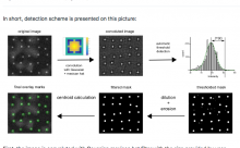
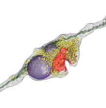
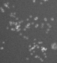
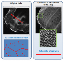
Nessys is a software written in Java for the automated identification of cell nuclei in biological images (3D + time). It is designed to perform well in complex samples, i.e when cells are particularly crowded and heterogeneous such as in embryos or in 3D cell cultures. Nessys is also fast and will work on large images which do not fit in memory.


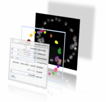
Nessys also offers an interactive user interface for the curation and validation of segmentation results. Think of this as a 3D painter / editor. This editor can also be used to generate manually segmented images to use as ground truth for testing the accuracy of the automated segmentation method.


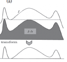
Finally Nessys, contains a utility for assessing the accuracy of the automated segmentation method. It works by comparing the result of the automated method to a manually generated ground truth. This utility will provide two types of output: a table with a number of metrics about the accuracy and an image representing a map of the mismatch between the result of the automated method and the ground truth.

has function
Description

3Dscript is a plugin for Fiji/ImageJ for creating 3D and 4D animations of microscope data. In contrast to existing 3D visualization packages, animations are not keyframe-based, but are described by a natural language-based syntax.

Description

Labkit is an open-source tool to segment truly large image data using sparse training data. It has an intuitive and responsive user interface based on Big Data Viewer, allowing users to conveniently browse and annotate even terabyte sized image volumes.

Update site: Labkit

has topic
need a thumbnail
Description

SciView is an ImageJ/FIJI plugin for 3D visualization of images and meshes. It uses the Scenery and ClearVolume infrastructure. SciView integrates ImageJ2 functionality, including ImageJ Ops and ImageJ Mesh, to provide the ability to interact with image and mesh data in 3D and interface with the popular Fiji software ecosystem.

An update site is available: http://sites.imagej.net/SciView/

has function
null
Description

NanoJ-SQUIRREL (Super-resolution Quantitative Image Rating and Reporting of Error Locations) is a software package designed for assessing and mapping errors and artefacts within super-resolution images. This is achieved through quantitative comparison with a reference image of the same structure (typically a widefield, TIRF or confocal image). SQUIRREL produces quantitative maps of image quality and resolution as well as global image quality metrics.

has function
SQUIRREL
Description

InspectJ is a free ImageJ/FIJI tool to inspect digital image integrity.

InspectJ_v2 is a newer version for advanced users. It applies additional features like histogram equalization and gamma correction for improved image inspections.

need a thumbnail
Description

OpenCV (Open Computer Vision) library for Icy. see more at http://opencv.org

has function
need a thumbnail
Description

NeuroMorph is a toolset designed to import, analyze, and visualize mesh models in Blender. It has been developed specifically for the morphological analysis of 3D objects derived from serial electron microscopy images of brain tissue, but much of its functionality can be applied to any 3D mesh. These mesh objects can be generated by any 3D image segmentation software, such as ilastik or Fiji

Description

This is a classical workflow for spot detection or blob like structures (vesicules, melanosomes,...)

Step 1 Laplacian of Gaussian to enhance spots . Paraeters= radius, about the average spot radius

Step 2 Detect minima (using Find Maxima with light background option to get minima). Parameter : Tolerance to Noise: to be tested, hard to predict. About the height of the enhanced feautures peaks

has topic
has function
spot detection
Description

The best way to start writing an ImageJ2 plugin (ImageJ2 developers call it command and not plugin) is to download the example command from github and modify it. There is a video tutorial on the whole workflow on how to do this on youtube.

has function
Description

This plugin detects a minimum cost z-surface in a 3D volume. A z surface is a topographic map indicating the altitude z as a function of the position (x,y) in the image. The cost of the surface depends on pixel intensity the surface is going through. This plugin find the z-surface with the lowest intensity in an image.

has function
Description

The interactive Watershed Fiji plugin provides an interactive way to explore local maxima and threshold values while a resulting label map is updated on the fly.

After the user has found a reliable parameter configuration, it is possible to apply the same parameters to other images in a headless mode, for example via ImageJ macro scripting.