Cell tracking

Description

CellStich proposes a set of tools for 3D segmentation from 2D segmentation: it reassembles 2D labels obtained from cell in slices in unique 3D labels across slices. It isparticularly robust to anisotropy, and is the ideal companion to cellpose 2D models or other 2D deep learning based models. One could also think about using it for cell tracking by overlap (using time as a third dimension).

cellstitch
Description

ELEPHANT is a platform for 3D cell tracking, based on incremental and interactive deep learning.
It implements a client-server architecture. The server is built as a web application that serves deep learning-based algorithms. The client application is implemented by extending Mastodon, providing a user interface for annotation, proofreading and visualization.

from https://elephant-track.github.io/#/v0.5/?id=_5-proofreading
Description

btrack is a Python library for multi object tracking, used to reconstruct trajectories in crowded fields. btrack implemented a residual U-Net model coupledd with a classification CNN to allow accurate instance segmentation of the cell nuclei. To track the cells over time and through cell divisions, btrack developed a Bayesian cell tracking methodology that uses input features from the images to enable the retrieval of multi-generational lineage information from a corpus of thousands of hours of live-cell imaging data.

need a thumbnail
Description

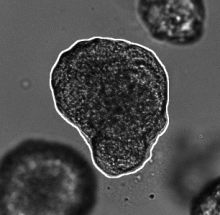
This workflow describes a deep-learning based pipeline for reliable single-organoid segmentation and tracking in 2D+t high-resolution brightfield microscopy of mouse mammary epithelial organoids. The pipeline involves a four-layer U-Net to infer semantic segmentation predictions, adaptive morphological filtering to establish candidate organoid instances, and a shape-similarity-constrained, instance-segmentation-correcting tracking step to associate the corresponding organoid instances in time.

It is particularly focused on automatically detecting an organoid located approximately in the center of the first frame and track all its subsequent instances in the remaining frames, emphasizing on accurate organoid boundary delineation. Furthermore, segmentation network was trained using plausible pix2pixHD-generated bioimage data. Syntheric image simulator code and data are also available here.

Adapted from https://cbia.fi.muni.cz/research/spatiotemporal/organoids.html
Description

TGMM is a cell tracking solution for large 3D volume (typically lightsheet).

It detects cell nuclei by fitting gaussians on their fluorescent intensity.

It can run on GPU using CUDA and is called via the command line.

has function
need a thumbnail
Description

OligoMacro Toolset, is an ImageJ macro-toolset aimed at isolating oligodendrocytes from wide-field images, tracking isolated cells, characterizing processes morphology along time, outputting numerical data and plotting them. It takes benefit of ImageJ built-in functions to process images and extract data, and relies on the R software in order to generate graphs.

need a thumbnail
Description


DeepCell is neural network library for single cell analysis, written in Python and built using TensorFlow and Keras.

DeepCell aids in biological analysis by automatically segmenting and classifying cells in optical microscopy images. This framework consumes raw images and provides uniquely annotated files as an output.

The jupyter session in the read docs are broken, but the one from the GitHub are functional (see usage example )

deepcell
Description

SIMPLETRACKER a simple particle tracking algorithm that can deal with gaps.

Tracking , or particle linking, consist in re-building the trajectories of one or several particles as they move along time. Their position is reported at each frame, but their identity is yet unknown: we do not know what particle in one frame corresponding to a particle in the previous frame. Tracking algorithms aim at providing a solution for this problem. 

simpletracker.m is - as the name says - a simple implementation of a tracking algorithm, that can deal with gaps. A gap happens when one particle that was detected in one frame is not detected in the subsequent one. If not dealt with, this generates a track break, or a gap, in the frame where the particle disappear, and a false new track in the frame where it re-appear. 

need a thumbnail
Description

MaMuT is an end user plugin that combines the BigDataViewer and TrackMate to provide an application that allow browsing, annotating and curating annotations for large image data.

Description

The purpose of the workflow is ....

First you need

need a thumbnail
Description

Summary

QuimP is software for tracking cellular shape changes and dynamic distributions of fluorescent reporters at the cell membrane. QuimP's unique selling point is the possibility to aggregate data from many cells in form of spatio-temporal maps of dynamic events, independently of cell size and shape. QuimP has been successfully applied to address a wide range of problems related to cell movement in many different cell types. 

Introduction

In transmembrane signalling the cell membrane plays a fundamental role in localising intracellular signalling components to specific sites of action, for example to reorganise the actomyosin cortex during cell polarisation and locomotion. The localisation of different components can be directly or indirectly visualised using fluorescence microscopy, for high-throughput screening commonly in 2D. A quantitative understanding demands segmentation and tracking of whole cells and fluorescence signals associated with the moving cell boundary, for example those associated with actin polymerisation at the cell front of locomoting cells. As regards segmentation, a wide range of methods can be used (threshold based, region growing, active contours or level sets) to obtain closed cell contours, which then are used to sample fluorescence adjacent to the cell edge in a straightforward manner. The most critical step however is cell edge tracking, which links points on contours at time t to corresponding points at t+1. Optical flow methods have been employed, but usually fail to meet the requirement that total fluorescence must not change. QuimP uses a method (ECMM, electrostatic contour migration method (Tyson et al., 2010) which has been shown to outperform traditional level set methods. ECMM minimises the sum of path lengths connecting all pairs of points, equivalent to minimising the energy required for cell deformation. The original segmentation based on an active contour method and outline tracking algorithms have been described in (Dormann et al., 2002; Tyson et al., 2010; Tyson et al., 2014).

Screenshot
Description

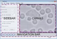
A standalone cell tracking software for single cell migration. Tracking of cells in tissue was also done in Drosophila germband.

GUI image (from http://sacan.biomed.drexel.edu/celltrack)
Description

## Short Summary Quote from the plugin page: >LineageTracker offers an ImageJ based framework which is easily extendible and has the capability to track cell lineages while being specifically designed to handle large cell displacements between frames. The methods are designed for fluorescent cells and have been used to analyse Schizosaccharomyces pombe, C2C12 mouse stem cells or migrating RPE cells. This tool also allows flexible cell segmentation and extendable in all aspects. The webpage is detailed with usage from ImageJ macro. Rather than being simply a component, the plugin is indeed a framework with set of components. ## Misc info A tip from the plugin author in ImageJ mailing list (08.Sep.2015): > We have an additional script to export only a selected range of frames. I can send you that if you think LineageTracker is something for you. To be on the safe side I would try it with an older version of ImageJ. We have experienced some problems, mostly related to Java. Java 8 seems to fix most of it. ## References 2630: Application example. 2631: Plugin Paper.

has function
need a thumbnail
Description

Quote: *A GUI-based program which manually detects spots and places them into previously detected meshes. Currently the program runs from MATLAB only. *

need a thumbnail
Description

This Matlab code demonstrates an edge-based active contour model as an application of the Distance Regularized Level Set Evolution (DRLSE) formulation.

initialisation
Description

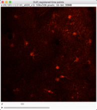
The workflow measures the growth of cells in 3D, combining an ImageJ macro for preprocessing and successive tracking using Imaris.  

The sample dataset (available in the github repository) contains 2-Photon images of neurons. The neurons were imaged in 3D at two time frames.To allow measuring significant differences in cell volume, the time gap between the frames is large (ca. 30 min) and the animal was removed in the waiting phase. For this reason, there is a considerable shift in sample position between the frames that has to be corrected before cell detection and tracking.

The workflow consists of following steps:

1. Import of single tiff slices [imageJ macro]

2. Organizing the data in a 4D time series with 2 time frames [imageJ macro]

3. Correction of shift between the time frames by rigid registration [imagJ macro]

4. Bleaching correction [imageJ macro]

5. Export of preprocessed image data in ics/ids format [imageJ macro]

6. Import of ics/ids data to Imaris [Imaris]

7. Cell object detection as "Imaris Surface Object" [Imaris]

8. Tracking cell objects over time [Imaris]

9. Split Tracks (use Imaris XT extension "Split Tracks") to generate single cell objects [Imaris]

10. Export the statistics: Select the complete folder, go to the statistics tab and use ‚Full Export’ [Imaris]

The preprocessing macro can be referenced here.

The sample images were acquired by Cordula Ulbrich (Petzold Group at German Center of Neurodegenerative Disesases (DZNE), Bonn, Germany).

Input data type: tiff

Output data type: data table

has function
Description

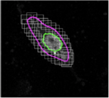
CellX is an open-source software package of workflow template for cell segmentation, intensity quantification, and cell tracking on a variety of microscopy images with distinguishable cell boundary.

Installation and step-by-step usage details are described in Mayer et al (2013). 

After users provide a few annotations of cell sizes and cell boundary profiles, it tries to match boundary profile pattern on cells thus provide segmentation and further tracking. It works the best on cells without extreme shapes and with a rather homogeneous boundary pattern. It may not work well on images with cells of sizes only a few pixels. Its output comprises control images for visual validation, text files for post-processing statistics, and MATLAB objects for advanced subsequent analysis.

Description

The QuimP software from Bretschneider group is deployed as ImageJ plugin and includes model-based cell segmentation, cell outline tracking and quantification of the spatially resolved speed of protrusions and retractions. The algorithm to calculate morphological dynamics is faster compared to other approaches (e.g. Machacek and Danuser, 2006). The reference paper describes the workflow for these analyses.

Description

Tracks a cell in a 2D video using active contours, and produces a list of ROI where intensity is measured and reported into a workbook. The cell must be first delineated with a ROI in the first image of the video.

need a thumbnail
Description

Oufti (previously named MicrobeTracker) is a MATLAB application / suite of tools for analysing fluorescent spots inside microbes. MicrobeTracker can identify cell outlines and fluorescent foci, and generate plots and statistics based on positions and intensity (kymographs, histograms etc.) The MATLAB code is easy to modify and extend to add additional plots and statistics: see e.g. Lesterlin et al. (2014).

The Outfi Forum is quite active.

Description

The goal of this workflow is to track cells captured in a time-lapse movie of a syncytial blastoderm stage Drosophila embryo and quantify their movement.

This example shows an example of object tracking. This pipeline analyzes a time-lapse experiment to identify the cells and track them from frame to frame, which is challenging since the cells are also moving. In addition, this pipeline also extracts metadata from the filename and uses groups the images by metadata in order to independently process several sequences of images and output the measurements of each.

Sample images

A portion of a time lapse movie of a syncytial blastoderm stage Drosophila embryo with a GFP-histone gene which renders chromatin fluorescent in live embryos. The movie shows nuclear divisions 10 through 13.

Victoria Foe made this movie on a Bio-Rad Radiance 2000 laser scanning confocal microscope using a 40X 1.3NA oil objective. The frames are 7 seconds apart and plays at 30 frames per second

GFP-histone transformed files provided by Rob Saint

V.Foe and G.Odell, . 26 July 2001

has function
Description

The workflow includes segmentation, tracking and quantifying morphological dynamics of moving cells in 3D. The authors have implemented the workflow in Matlab, but so far there is no download link provided. To apply this workflow, we recommend to contact the authors or to implement the worflow based on the detailed description in the original paper.

has function
need a thumbnail
Description

u-track is a multiple-particle tracking Matlab software that is designed to (1) track dense particle fields, (2) close gaps in particle trajectories resulting from detection failure, and (3) capture particle merging and splitting events resulting from occlusion or genuine aggregation and dissociation events. Its core is based on formulating correspondence problems as linear assignment problems and searching for a globally optimal solution.

Data can be read using bio-format and interfaced with OMero data base.

It comes as a standalone software, but can be used as a library, which is according to the authors the most widely used version of it.

  • Version 2.2 adds parallel processing functionality for multi-movie datasets when using the GUI.
  • Version 2.1 enables the analysis of movies stored on an OMERO server
  • Version 2.0 includes two new tracking applications: microtubule plus-end tracking (previously distributed as plusTipTracker) and nuclei tracking
  • A third optional processing step has been added to the analysis workflow, track analysis, with two methods: motion analysis and microtubule plus-end classification

For more information, please see Jaqaman et al., Nature Methods 5, pp. 695-702 (2008). Besides basic particle tracking, the software supports the features described in Applegate et al. J. Struct. Biol. 176(2):168-84. 2011 for tracking microtubule plus end markers; and in Ng et al. J. Cell Biol. 199(3):545-63. 2012 for tracking fluorescently-labeled cell nuclei.


Description

CellTracker software is a platform for tracking nuclear and cytoplasmic fluorescence intensities from live cell microscopy time series data.


Requires visual C++

Description

Dedicated to score tracking dataset from the ISBI tracking challenge

has function
Description

"This ImageJ plugin (CGE) is a semi-automatic tool to detect and track moving cell, and to measure the fluorescent protein expression level. CGE extracts the trajectory of the cells by tracking their displacements, makes the delineation of cell nucleus or whole cell, and finally yields measurements of various features, like reporter protein expression level, cell displacement."

has function