ImageJ Macros

Description

The BioVoxxel Toolbox is a suite which contains plugins and some macros dealing with image filtering, image segmentation and binary image processing and analysis. The following plugins are hosted here:

  • Extended Particle Analyzer
  • Binary Feature Extractor
  • Speckle Inspector
  • Watershed Irregular Features
  • EDM Binary Operations
  • Filter Check
  • Pseudo flat-field correction
  • Convoluted Background Subtraction
  • 2D Particle Distribution (Distribution_Analysis)
  • Cluster Indicator
  • SSIDC Cluster Indicator
  • Gaussian weighted Median filter
  • Adaptive Filter
  • Enhance True Color Contrast
  • Mode and Differential Limited Mean Binarization
  • Basic Recursive Filter
has topic
Description

Fast4DReg is a Fiji macro for drift correction for 2D and 3D video and is able to correct drift in all x-, y- and/or z-directions. Fast4DReg creates intensity projections along both axes and estimates their drift using cross-correlation based drift correction, and then translates the video frame by frame. Additionally, Fast4DReg can be used for alignment multi-channel 2D or 3D images which is particularly useful for instruments that suffer from a misalignment of channels.

has function
Description

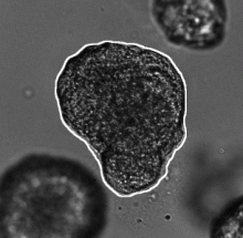
This workflow describes a deep-learning based pipeline for reliable single-organoid segmentation and tracking in 2D+t high-resolution brightfield microscopy of mouse mammary epithelial organoids. The pipeline involves a four-layer U-Net to infer semantic segmentation predictions, adaptive morphological filtering to establish candidate organoid instances, and a shape-similarity-constrained, instance-segmentation-correcting tracking step to associate the corresponding organoid instances in time.

It is particularly focused on automatically detecting an organoid located approximately in the center of the first frame and track all its subsequent instances in the remaining frames, emphasizing on accurate organoid boundary delineation. Furthermore, segmentation network was trained using plausible pix2pixHD-generated bioimage data. Syntheric image simulator code and data are also available here.

Adapted from https://cbia.fi.muni.cz/research/spatiotemporal/organoids.html
Description

It stitches 3D tiles from terabyte-size microscopy datasets. Stitching does not require any prior information on the actual positions of the tiles, sample fiducials, or conversion of raw TIFF images, and the stitched images can be explored instantly.

MosaicExplorerJ was specifically designed to process lightsheet microscopy datasets from optically cleared samples. It can handle multiple fluorescence channels, dual-side lightsheet illumination and dual-side camera detection.

GPU-Accelerating ImageJ Macro Image Processing Workflows Using CLIJ

Submitted by czhang on Thu, 04/27/2023 - 09:55

This chapter is part of this book. The chapter introduces GPU-accelerated image processing in ImageJ/Fiji. The reader is expected to have some pre-existing knowledge of ImageJ Macro programming. Core concepts such as variables, for-loops, and functions are essential. The chapter provides basic guidelines for improved performance in typical image processing workflows.

Description

The authors present an ImageJ-based, semi-automated phagocytosis workflow to rapidly quantitate three distinct stages during the early engulfment of opsonized beads.

Customizing ImageJ

These slides give an introduction to user interfacre customization in ImageJ using ImageJ Macro and to ImageJ Macro Markdown.

Big thanks to Jerome Mutterer (IBMP) and Nicolas De Francesco (IMBICE) who shared material openly I reused when making these slides.

Supplementary material is available as well under this doi: 10.5281/zenodo.4561714

Description

The tool exports rectangular regions, defined with the NDP.view 2 software (hammatsu) from the highest resolution version of the ndpi-images and saves them as tif-files.

Click the button and select the input folder. The input folder must contain pairs of ndpi and ndpa files. The regions will be exported to a subfolder of the input folder names zones.

has topic
has function
imagej toolset to export regions from ndpi and ndpa-files
Description

The tool allows to measure the area of the invading spheroïd in a 3D cell invasion assay. It can also count and measure the area of the nuclei within the spheroïd.

need a thumbnail
Description

This tool allows to analyze morphological characteristics of complex roots. While for young roots the root system architecture can be analyzed automatically, this is often not possible for more developed roots. The tool is inspired by the Sholl analysis used in neuronal studies. The tool creates a binary mask and the Euclidean Distance Transform from the input image. It then allows to draw concentric circles around a base point and to extract measures on or within the circles. Instead of circles, which present the distance from the base point, horizontal lines can be used, which present the distance in the soil from the base-line. The following features are currently implemented:

  • The area of the root per distance/depth.
  • The number of border pixel per distance/depth, giving an idea of the surface in contact with the soil.
  • The maximum radius per distance/depth of a root, measured at the crossing points with the circles or lines.
  • The number of crossings of roots with the circles or lines.
  • The maximum distance to the left and the right from the vertical axis at crossing points with the circles or lines.
Concentric circles on the mask of a root, created by the Analyze Complex Roots Tool
Description

Analyze the clustering behavior of nuclei in 3D images. The centers of the nuclei are detected. The nuclei are filtered by the presence of a signal in a different channel. The clustering is done with the density based algorithm DBSCAN. The nearest neighbor distances between all nuclei and those outside and inside of the clusters are calculated.

has function
Description

This macro toolset offers additional click tools for the rapid annotations of ROI in ImageJ/Fiji.

The ROI 1-click tools can be setup with a predefined shape, and custom actions to perform upon click (Add to ROI Manager, Run Measure, Go to next slice, run a macro command...)

To install in Fiji, just activate the ROI 1-click tools 

Description

Blood vessels tracing in 3D image from 3D Gaussian blurring (user defined radius), local thresholding (user defined radius and offset) and 3D skeletonization. Dockerized version for BIAFLOWS,

need a thumbnail
Description

Blood vessels tracing in 3D image from Tubeness filtering (user defined scale), 3D opening (radius set to 2), thresholding (user defined level) and 3D skeletonization.

need a thumbnail
Description

Object tracking. For each time-frame, an image mask is obtained from median filtering (user defined radius), thresholding (user defined level) and hole filling. Convex objects are split apart by distance map watershed from regional intensity maxima (user defined noise tolerance), eroded (user defined radius) and analyzed as 3D particles (assuming some overlap between objects from a frame to the next frame). Finally, division events are analyzed and accounted for to relabel objects.

has function
need a thumbnail
Description

Particle tracking in 2D time-lapse based on linking closest regional intensity minima (user defined noise tolerance) detected from Laplacian of Gaussian filtered images (user defined radius). A maximum linking distance is set (user defined).

has function
need a thumbnail
Description

The macro will segment nuclei and separate clustered nuclei in a 3D image using a 2D Gaussian blur, followed by Thresholding, 2D hole filling and a 2D watershed. As a result an index-mask image is written for each input image.

need a thumbnail
Description

3D spot detection using the Determinant of Hessian (DoH) and the detection of 3D minima.

need a thumbnail
Description

This workflow detects spots in a 2D image by filtering the image by Laplacian of Gaussian (user defined radius), thresholding (user defined threshold) and finding local intensity maxima in mask distance map (Dmap).

need a thumbnail
Description

OligoMacro Toolset, is an ImageJ macro-toolset aimed at isolating oligodendrocytes from wide-field images, tracking isolated cells, characterizing processes morphology along time, outputting numerical data and plotting them. It takes benefit of ImageJ built-in functions to process images and extract data, and relies on the R software in order to generate graphs.

need a thumbnail
Description

We have developed WormScan, an automated image acquisition system that allows quantitative analysis of each of these four phenotypes on standard NGM plates seeded with E. coli. This system is very easy to implement and has the capacity to be used in high-throughput analysis.

Description

Protein array is used to analyze protein expressions by screening simultaneously several protein-molecule interactions such as protein-protein and protein-DNA interactions. In most cases, the detection of interactions leads to an image containing numerous lines of spots that will be analyzed by comparing tables of intensity values. To describe the observed different patterns of expression, users generally show histograms with the original associated images [1]. The “Protein Array Analyzer” gives a friendly way to exploit this type of analysis, thus allowing quantification, image modeling and comparative analysis of patterns.

The Protein Array Analyzer, which was programmed in ImageJ’s macro language, is an extention of the Dot Blot Analyzer, [2], [3] a graphically interfaced tool that greatly simplifying analysis of dot arrays.