Image thresholding

Synonyms
Thresholding
Clustering-based image thresholding
Description

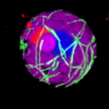
VTK is an open-source software system for image processing, 3D graphics, volume rendering and visualization. VTK includes many advanced algorithms (e.g., surface reconstruction, implicit modeling, decimation) and rendering techniques (e.g., hardware-accelerated volume rendering, LOD control).

VTK is used by academicians for teaching and research; by government research institutions such as Los Alamos National Lab in the US or CINECA in Italy; and by many commercial firms who use VTK to build or extend products.

The origin of VTK is with the textbook "The Visualization Toolkit, an Object-Oriented Approach to 3D Graphics" originally published by Prentice Hall and now published by Kitware, Inc. (Third Edition ISBN 1-930934-07-6). VTK has grown (since its initial release in 1994) to a world-wide user base in the commercial, academic, and research communities.

Description

MiNA is a simplified workflow for analyzing mitochondrial morphology using fluorescence images or 3D stacks in Fiji. The workflow makes use of ImageJ Ops3D ViewerSkeletonize (2D/3D)Analyze Skeleton, and Ridge Detection. In short, the tool estimates mitochondrial footprint (or volume) from a binarized copy of the image as well as the lengths of mitochondrial structures using a topological skeleton. The values are reported in a table and overlays (or a 3D rendering) are generated to assess the accuracy of the analysis.

example skeleton image (from https://imagej.net/plugins/mina#processing-pipeline-and-usage)
Description

 This ImageJ function automatically or interactively sets lower and upper threshold values, segmenting grayscale images into features of interest and background.

has function
need a thumbnail
Description

QuantiFish is a quantification program intended for measuring fluorescence in images of zebrafish, although use with images of other specimens is possible. This package is geared towards analysis of fluorescent infection models. The software is designed to automate processing of images of single fish, and outputs results as a .csv file. Alongside measures of total fluorescence above a threshold, this package also introduces several measures for dissemination and distribution of fluorescence throughout the specimen.

QuantiFish User Interface
Description

CLIJ2 is a GPU-accelerated image processing library for ImageJ/FijiIcy, Matlab and Java. It comes with hundreds of operations for filteringbinarizinglabelingmeasuring in images, projectionstransformations and mathematical operations for images. While most of these are classical image processing operations, CLIJ2 also allows performing operations on matrices potentially representing neighborhood relationships between cells and pixels.

CLIJ2 was developed to process images from fluorescence microscopy data of developing cells, tissues, organoids and organisms.

Description

The MIPAV (Medical Image Processing, Analysis, and Visualization) application enables quantitative analysis and visualization of medical images of numerous modalities such as PET, MRI, CT, or microscopy. Using MIPAV's standard user-interface and analysis tools, researchers at remote sites (via the internet) can easily share research data and analyses, thereby enhancing their ability to research, diagnose, monitor, and treat medical disorders.

Description

The ultimate goal of the NET framework is to make images of networks processable by computers. Therefore we want to have a pixel based image as input, as output we want a representation of the network visible in the image that retains as much information about the original network as possible. NET achives this by first segmenting the image and then vectorizing the network and then extracting information. The information we extract is

  • First and foremost the graph of the network. We find the crossings (nodes) and connections between crossings (edges) and therefore extract information about the neighborhood relations, the topology of the network.
  • We also extract the coordinates of all nodes which enables us to embed them into space. We therefore extract information about the geometry of the network.
  • Last but not least we track the radii of the edges in the extraction process. Therefore every edge has a radius which can be identified with its conductivity.

In the following we will first provide detailed instructions on how to install NET on several platforms. Then we describe the functionality and options of each of the four scripts that make up the NET framework.

has topic
need a thumbnail
Description

"we present a new fully automated 3D reconstruction algorithm, called TReMAP, short for Tracing, Reverse Mapping and Assembling of 2D Projections. Instead of tracing a 3D image directly in the 3D space as seen in majority of the tracing methods, we first trace the 2D projection trees in 2Dplanes, followed by reverse-mapping the resulting 2D tracing results back into the 3D space as 3D curves; then we use a minimal spanning tree (MST) method to assemble all the 3D curves to generate the final 3D reconstruction. Because we simplify a 3D reconstruction problem into 2D, the computational costs are reduced dramatically." 

Suitable for high throughput neuron image analysis (image sizes >10GB). This plugin can be used with default parameters or user-defined parameters.

Example_TReMAP_Result
Description

"We have developed an automatic graph algorithm, called the all-path pruning (APP), to trace the 3D structure of a neuron. To avoid potential mis-tracing of some parts of a neuron, an APP first produces an initial over-reconstruction, by tracing the optimal geodesic shortest path from the seed location to every possible destination voxel/pixel location in the image. Since the initial reconstruction contains all the possible paths and thus could contain redundant structural components (SC), we simplify the entire reconstruction without compromising its connectedness by pruning the redundant structural elements, using a new maximal- covering minimal-redundant (MCMR) subgraph algorithm. We show that MCMR has a linear computational complexity and will converge. We examined the performance of our method using challenging 3D neuronal image datasets of model organisms (e.g. fruit fly)"

This plugin can be used with default parameters or user-defined parameters.

APP_Vaa3D_example_results
Description

This is an ImageJ plugin to analyze bacterial cells. It provides a user-friendly interface and a powerful suite of detection, analysis and data presentation tools. It works with individual phase or fluorescence images as well as stacks, hyperstacks, and folders of any of these types. Even large image sets are analyzed rapidly generating raw tabular data that can either be saved or copied as is, or have additional statistical analysis performed and graphically represented directly from within MicrobeJ, making it an all-in-one image analysis solution.

need a thumbnail
Description

Adiposoft is an automated Open Source software for the analysis of adipose tissue cellularity in histological sections.

Example data can be found on the plugin description page in ImageJ wiki (download link). There is also a link to a MATLAB version of the workflow.

has topic
Description

The Measure Rosette Area Tool allows to measure the area of the rosettes of arabidopsis plants.

See: http://dev.mri.cnrs.fr/projects/imagej-macros/wiki/Measure_Rosette_Area…

Example data: http://biii.eu/node/1146http://biii.eu/node/1145

has function
Description

This method allows to compute a threshold that preserves the moments of an image. In ImageJ/Fiji, you can access it in Image->Ajust->Threshold and choose Moments in the list. In Aphelion, the tool is in Segmentation->Threshold->AphImgMomentThreshold The original paper is 2449

need a thumbnail
Description

Simple workflow description for ImageJ, step-by-step description for delineating focal adhesions, count and characterize their positions.  

Measurement of dynamics is not involved.

Description

The tool measures the total length of the microtubules in a 3D image.

See: http://dev.mri.cnrs.fr/projects/imagej-macros/wiki/Microtubules_Tool_(3…

You can find a test image here.

3D microtubules
Description

The Arabidopsis Seedlings Tool allows to analyze the germination and seedling growth of Arabidopsis (Arabidopsis thaliana) in liquid culture. It measures the surface of green pixels per well in images containing multiple wells. It can be run in batch mode on a series of images. It writes a spreadsheet file with the measured area per well and saves a control image showing the green surface that has been detected per well. 

See http://dev.mri.cnrs.fr/projects/imagej-macros/wiki/Arabidopsis_Seedlings_Tool

Test images can be found here.

has function
ImageJ toolbar of the arabidopsis seedlings tool

ITK

Description

ITK is an open-source, cross-platform system that provides developers with an extensive suite of software tools for image analysis.

Developed through extreme programming methodologies, ITK employs leading-edge algorithms for registering and segmenting multidimensional data. It is widely used and contributed in the medical imaging field.

Strengths

Highly optimized C++, well commented Consistently updated (new) algorithms many tools and softwares are built upon it connected with VTK Insight Journal (open code and sample data) Extensive list of examples & tutorials

Limitations

yet detached from the bioimage analysis world hard to use for end users without development skills

itk
Description

This library gives the numpy-based infrastructure functions for image processing with a focus on bioimage informatics. It provides image filtering and morphological processing as well as feature computation (both image-level features such as Haralick texture features and SURF local features). These can be used with other Python-based libraries for machine learning to build a complete analysis pipeline.

Mahotas is appropriate for users comfortable with programming or builders of end-user tools.

==== Strengths

The major strengths are in speed and quality of documentation. Almost all of the functionality is implemented in for multiple dimensions. It can be used with other Python packages which provide additional functionality.

Mahotas and all packages on which it relies are open-source.

Description

This plugin threshold an image using the Maximum Entropy algorithm, which aims at maximizing the inter-class entropy. Entropy is defined as -sum(p.*log2(p)), where p contains the histogram bin counts. This thresholding is very useful to segment images with few bright objects on large dark background. In ImageJ/FIJI you can acces this tool in Image->Adjust->Threshold and choose in the list In Aphelion, you can access this tool in Seglmentation->Threshold-> AphImgEntropyThreshold

need a thumbnail
Description

This plugin automatically threshold an image using the Mixture Modeling algorithm. It is an histogram-based technique that assumes that the histogram distribution is represented by two Gaussian curves.

has function
need a thumbnail
Description

This ImageJ plugin segments the image in classes by thresholding. It uses the same algorithm found in Otsu Thresholding, but was adapted to output more than 2 classes out of the process.

has function
need a thumbnail
Description

The method to use for thresholding. Currently only OTSU is available.

has function
need a thumbnail
Description

Bundled with Fiji. "Do all" is the great feature...

has function
Description

Extract image features based on one or more intensity thresholds, and output the result as a labeled image or as a region of interest.

has function
need a thumbnail
Description

For each ROI, computes the number of pixel over a threshold. This plugin also provides the density and outputs results as an excel file.

has topic
need a thumbnail