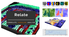
Scanning electron microscopy

Description


Relate is a correlative software package optimised to work with EM, EDS, EBSD, & AFM data and images.  It provides the tools you need to correlate data from different microscopes, visualise multi-layered data in 2D and 3D, and conduct correlative analyses.

  • Combining data from different imaging modalities (e.g. AFM, EDS & EBSD)

  • Interactive display of multi-layer correlated data

  • Analytical tools for metadata interrogation

  • Documented workflows and processes

Correlate

  • Import data from AZtec using the H5oina file format
  • Import AFM data
  • Correlate both sets of data using intuitive image overlays and image matching tools
  • Produce combined multimodal datasets

Visualise

  • 2D display of multi-layered data
  • 3D visualisation of topography combined with AFM material properties, EM images, and EDS & EBSD map overlays
  • Customisation of colour palettes, data overlays, image rendering options, and document display
  • Export images and animations

Analyse

  • Generate profile (cross section) views of multimodal data
  • Measure and quantify data across multiple layers
  • Analyse areas via data thresholding using amount of x-ray counts, phase maps, height, or other material properties.
  • Select an extensive range of measurement parameters
  • Export analytical data to text or CSV files
Relate analysis workflow example
Description

A collection of Java tools and HTTP services (APIs) for rendering transformed image tiles that includes:

The basic concept is to render images (tiles) based on transformation files, without having to store the big generated image from an alignment of tiles (mosaicking).

Description

SuRVoS: Super-Region Volume Segmentation workbench

A volume is first partitioned into Super-Regions (superpixels or supervoxels) and then interactively segmented by the user providing training annotations. SuRVoS can then learn from and extend the annotations to the whole volume.

User interface of SuRVoS showing example annotation on soft x-ray tomography data
Description

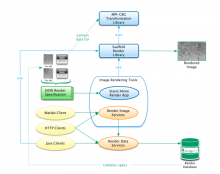
By combining multiple image alignment and tracing into one program, Reconstruct (TM) allows images to be processed more efficiently. Tracing can be done directly on the transformed images and alignments can be asily modified. Reconstruct (TM) was developed from years of experience working with high magnification serial section images of brain tissue. (Extracted from User Manual)

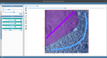
"The original platform of the Reconstruct program allows a user to trace objects in serial sections by manually drawing the outline of each object on each section, which is time-consuming. We modified Reconstruct to enable semi-automatic tracing of axons using a region-growing algorithm called wildfire."

Reconstruct_standaloneapp_example_Results