Commercial

Description

MetaXpress or in full name "MetaXpress® High-Content Image Acquisition and Analysis Software" is a commercially available closed source software for high-content analysis from Molecular Devices, LLC.. The program is a kind of visually guided workflow programming environment. There is a programming module called CME (custom module editor) which lets one setup integrated workflows for bioimage analysis with visual feedback. It is designed for high-throughput in connection with a included database which stores the experimental data. 

It has several toolboxes for semiautomated processing of various tasks:

3D Analysis (requires Custom Module Editor), Curve fitting, Transmitted light segmentation (requires Custom Module Editors), Angiogenesis tube formation, Cell cycle, Cell health, Cell scoring , Count nuclei, Granularity, Live/dead , Mitotic index, Micronuclei , Monopole detection, Multi-Wavelength cell scoring, Multi-wavelength translocation, Neurite outgrowth , Transfluor® Assay, Translocation* (includes Translocation-Enhanced*) , Transfluor HT Assay , Nuclear translocation HAT, Cell proliferation HT

After the workflow is setup it is possible to apply it automatically to a stack of stored images. The derived data from those analyses is stored in the metaxpress database and can be exported from there.

The use of each toolbox requires a separate license.

Description
# Install the ultralytics package from PyPI
pip install ultralytics

You can also install ultralytics directly from the Ultralytics GitHub repository. This can be useful if you want the latest development version. Ensure you have the Git command-line tool installed, and then run:

# Install the ultralytics package from GitHub
pip install git+https://github.com/ultralytics/ultralytics.git@main
Description

Ultralytics creates cutting-edge, state-of-the-art (SOTA) YOLO models built on years of foundational research in computer vision and AI. Constantly updated for performance and flexibility, our models are fast, accurate, and easy to use. They excel at object detection, tracking, instance segmentation, image classification, and pose estimation tasks.

Description

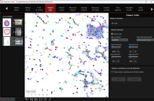
HistoMetriX is a quantitative histology and digital pathology image analysis software designed for biomedical research. It enables automated and AI-assisted analysis of brightfield and fluorescence microscopy images, including whole-slide images and tissue microarrays.

HistoMetriX combines deep-learning-based tissue segmentation with cell detection and classification, quantitative morphology and intensity measurements, biomarker quantification, and spatial analysis. Custom AI models can be trained to identify tissue structures and biological phenotypes specific to a study.

The software provides interactive visualization and validation of analysis results and is designed to support reproducible quantitative workflows for applications including oncology, immunology, dermatology, toxicology and biomedical research. Research use only.

HistoMetriX software for AI-powered histology and digital pathology image analysis
Description

The Incucyte® Base Analysis Software provides a guided interface and purpose-built tools, which include the process of acquiring, viewing, analyzing and sharing images of living cells.

need a thumbnail