Free and open source

Description

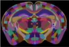
A python based tool for registering a template brain (from the Allen Reference Atlas) to the sample image, an extension of aMAP. Once this is complete, any other image in the template space can be aligned with the sample (such as region annotations, for segmentation of the sample image). The template to sample transformation can also be inverted, allowing sample images to be aligned in a common coordinate space. Current registration backend is niftyreg.


Component of BrainGlobe Initiative

Description

BraiAn is an open-source suite of tools designed to simplify signal quantification, analysis and visualization of large datasets typically obtained in whole-brain imaging experiments, following registration to an atlas. 

The package consists of two separate modules.

  1. BrainAnDetect: A QuPath extension for multi-channel cell segmentation across large and variable datasets. It leverages QuPath's built in algorithms for cell detection, and features additional options for refining signal quantification, including machine-learning-based object classification, region-specific cell segmentation, multiple marker co-expression analysis, and an interface for selective exclusion of damaged tissue portions.
  2. BraiAnalyse: A modular Python library for the easy navigation, visualization, and analysis of whole-brain quantification outputs.
has topic
need a thumbnail
Description

Ultrack is a versatile and scalable cell tracking method designed to address the challenges of tracking cells across 2D, 3D, and multichannel timelapse recordings, especially in complex and crowded tissues where segmentation is often ambiguous. By evaluating multiple candidate segmentations and employing temporal consistency, Ultrack ensures robust performance under segmentation uncertainty. Ultrack's methodology is explained here.

(from https://github.com/royerlab/ultrack)

Description

A generalist framework for multi-dimensional automatic spot detection and quantification.

SpotMAX is designed to accomplish two tasks:

  1. Detecting and quantifying globular-like structures (a.k.a. "spots")
  2. Segmenting and quantifying fluorescently labelled structures

It supports 2D, 3D, 4D, and 5D data, i.e., z-stacks, timelapse, and multiple fluorescence channels (and combinations thereof).

has function
SpotMAX Logo
Description

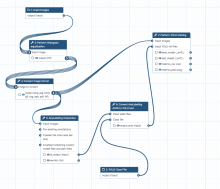
This workflow is the integration of YOLO (You Only Look Once) machine learning models, image pre-processing scripts and labeling tools within the Galaxy platform. Galaxy is an open, web-based platform used primarily for data analysis in computational biology, but it also has applications in image processing and other fields. 

How the Galaxy YOLO image segmentation tool works

The combination of Galaxy and YOLO allows researchers to perform object detection and image analysis without requiring extensive programming knowledge. Here's how it generally works: 

  • Web-based interface: Galaxy provides a graphical, user-friendly interface to access powerful analysis tools. Users can simply upload their image data, select the YOLO tool, and run the analysis.
  • YOLO model execution: The Galaxy tool executes a pre-trained YOLO model, often from the Ultralytics framework, on the input images. These models can perform tasks like object detection (drawing bounding boxes) or instance segmentation (creating pixel-level masks).
  • Training and prediction: Some tools allow for both model training and prediction. Users can train a custom YOLO model on their own labeled datasets to detect specific objects of interest. For example, bioimage analysis may involve detecting cells or other structures.
  • Other integrations: Other machine-learning tools can be integrated with YOLO in Galaxy. For instance, the AnyLabeling tool supports YOLO for semi-automated and active learning-based data annotation.