Bioimage informatics

Synonyms
Bioimage analysis
Description

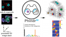
Phindr3D is a comprehensive shallow-learning framework for automated quantitative phenotyping of three-dimensional (3D) high content screening image data using unsupervised data-driven voxel-based feature learning, which enables computationally facile classification, clustering and data visualization.

Please see our GitHub page and the original publication for details.

Description

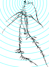
This tool allows to analyze morphological characteristics of complex roots. While for young roots the root system architecture can be analyzed automatically, this is often not possible for more developed roots. The tool is inspired by the Sholl analysis used in neuronal studies. The tool creates a binary mask and the Euclidean Distance Transform from the input image. It then allows to draw concentric circles around a base point and to extract measures on or within the circles. Instead of circles, which present the distance from the base point, horizontal lines can be used, which present the distance in the soil from the base-line. The following features are currently implemented:

  • The area of the root per distance/depth.
  • The number of border pixel per distance/depth, giving an idea of the surface in contact with the soil.
  • The maximum radius per distance/depth of a root, measured at the crossing points with the circles or lines.
  • The number of crossings of roots with the circles or lines.
  • The maximum distance to the left and the right from the vertical axis at crossing points with the circles or lines.
Concentric circles on the mask of a root, created by the Analyze Complex Roots Tool
Description

Quote:

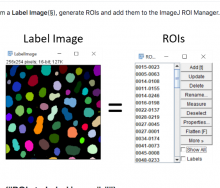
LaRoME = Label + Region Of Interest + Measure

Label image (aka Count Masks): An image in which pixels of an object have all the same value. Each object has a unique value.

Measurement image: An image in which pixels of an object have all the same value, corresponding to a measurement (Area, Angle, Mean...)

has function

Data Science in Cell Imaging (DSCI) course material

Submitted by assafzar on Thu, 12/31/2020 - 10:08

Graduate course at the department of Software and Information Systems Engineering at Ben-Gurion University of the Negev.

The recent explosion in high-content, dynamic and multidimensional imaging data is transforming cell imaging into a “Data Science” field. This course will review the state-of-the-art in visualizing, processing, integrating and mining massive cell image data sets, deciphering complex patterns and turning them into new biological insight. It will include a mix of approaches in computer science, machine learning and computer vision applied to bio-imaging data.

Description

Set of KNIME workflows for the training of a deep learning model for image-classification with custom images and classes.

The workflows take ground-truth category annotations as a table generated by the qualitative annotations plugins in Fiji.

Workflows for the training of a model AND for the prediction of image-category for new images are provided.

There are different workflows if you do:

- binary image-classification (images get classified in 1 category out of 2 possible categories) 

- classification from possibly more than 2 categories (images are classified in 1 category out of N possible categories).

The training workflows take care of image pre-processing and allows the visualization of the training and validation losses in real time along the training.  

For the training, transfer learning from a pre-trained VGG16 base is performed, with freshly initialized fully connected layers.

Only the fully connected layers are trained, the VGG16 base is frozen is this workflow, but once the fully connected layers trained the base could also be finetuned. In practice, it often works well with the frozen base.

has function