Component

A Component is an implementation of certain image processing / analysis algorithms.

Each component alone does not solve a Bioimage Analysis problem.

These problems can be addressed by combining such components into workflows.

Description

NODeJ is an ImageJ plugin for 3D segmentation of nuclear objects.

"The three-dimensional nuclear arrangement of chromatin impacts many cellular processes operating at the DNA level in animal and plant systems. Chromatin organization is a dynamic process that can be affected by biotic and abiotic stresses. Three-dimensional imaging technology allows to follow these dynamic changes, but only a few semi-automated processing methods currently exist for quantitative analysis of the 3D chromatin organization. We present an automated method, Nuclear Object DetectionJ (NODeJ), developed as an imageJ plugin. This program segments and analyzes high intensity domains in nuclei from 3D images. NODeJ performs a Laplacian convolution on the mask of a nucleus to enhance the contrast of intra-nuclear objects and allow their detection. We reanalyzed public datasets and determined that NODeJ is able to accurately identify heterochromatin domains from a diverse set of Arabidopsis thaliana nuclei stained with DAPI or Hoechst. NODeJ is also able to detect signals in nuclei from DNA FISH experiments, allowing for the analysis of specific targets of interest. NODeJ allows for efficient automated analysis of subnuclear structures by avoiding the semi-automated steps, resulting in reduced processing time and analytical bias. NODeJ is written in Java and provided as an ImageJ plugin with a command line option to perform more high-throughput analyses. NODeJ can be downloaded from https://gitlab.com/axpoulet/image2danalysis/-/releases with source code, documentation and further information avaliable at https://gitlab.com/axpoulet/image2danalysis . The images used in this study are publicly available at https://www.brookes.ac.uk/indepth/images/ and https://doi-org.osaka-u.idm.oclc.org/10.15454/1HSOIE ."

has function
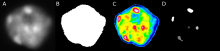
A DAPI-stained nucleus at left, followed by a white segmentation mask, a false-color heatmap, and segmented heterochromatin blocks.
Description

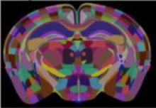
A python based tool for registering a template brain (from the Allen Reference Atlas) to the sample image, an extension of aMAP. Once this is complete, any other image in the template space can be aligned with the sample (such as region annotations, for segmentation of the sample image). The template to sample transformation can also be inverted, allowing sample images to be aligned in a common coordinate space. Current registration backend is niftyreg.


Component of BrainGlobe Initiative

Description

A generalist framework for multi-dimensional automatic spot detection and quantification.

SpotMAX is designed to accomplish two tasks:

  1. Detecting and quantifying globular-like structures (a.k.a. "spots")
  2. Segmenting and quantifying fluorescently labelled structures

It supports 2D, 3D, 4D, and 5D data, i.e., z-stacks, timelapse, and multiple fluorescence channels (and combinations thereof).

has function
SpotMAX Logo
Description

Tools for segmentation and tracking in microscopy build on top of Segment Anything. Segment and track objects in microscopy images interactively with a few clicks.

uSAM logo
Description

This is a placeholder component created for demonstration purposes. It serves as an example of how to create a minimal software entry in BIII. This component represents a basic image processing algorithm that can be used as part of a bioimage analysis workflow.

need a thumbnail