Image deconvolution

Description

In light-sheet microscopy, overall image content and resolution are improved by acquiring and fusing multiple views of the sample from different directions. State-of-the-art multi-view (MV) deconvolution simultaneously fuses and deconvolves the images in 3D, but processing takes a multiple of the acquisition time and constitutes the bottleneck in the imaging pipeline. Here, we show that MV deconvolution in 3D can finally be achieved in real-time by processing cross-sectional planes individually on the massively parallel architecture of a graphics processing unit (GPU). Our approximation is valid in the typical case where the rotation axis lies in the imaging plane.

need a thumbnail
Description

The Multiview Reconstruction software package enables users to register, fuse, deconvolve and view multiview microscopy images. The software is designed for lightsheet fluorescence microscopy (LSFM), but is applicable to any form of three or higher dimensional imaging modalities like confocal timeseries or multicolor stacks. 

need a thumbnail
Description

The BigStitcher is a software package that allows simple and efficient alignment of multi-tile and multi-angle image datasets, for example acquired by lightsheet, widefield or confocal microscopes. The software supports images of almost arbitrary size ranging from very small images up to volumes in the range of many terabytes, which are for example produced when acquiring cleared tissue samples with lightsheet microscopy.

Description

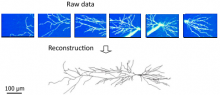
ORION: Online Reconstruction and functional Imaging Of Neurons: segmentation and tracing of neurons for reconstruction.

A project to develop tools that explore single neuron function via sophisticated image analysis. ORION software bridges advanced optical imaging and compartmental modeling of neuronal function by rapidly, accurately, and robustly generating, from structural image data, a cylindrical morphology model suitable for simulating neuronal function. The goal of this project is to develop a computational and experimental framework to allow real-time mapping of functional imaging data (e.g., spatio-temporal patterns of dendritic voltages or intracellularions) to neuronal structure, during the very limited duration of an acute experiment.

ORION_example_result
Description

SRRF is a high-performance analytical approach for Live-cell Super-Resolution Microscopy, provided as a fast GPU-enabled ImageJ plugin. SRRF is capable of extracting high-fidelity super-resolution information from TIRF, widefield and confocals using conventional fluorophores such as GFP. SRRF is capable of live-cell imaging over timescales ranging from minutes to hours.

Comparison TIRF - SRRF