neurites

SNEMI3D: 3D Segmentation of neurites in EM images

Submitted by Perrine on Mon, 03/06/2023 - 16:45

In this challenge, a full stack of electron microscopy (EM) slices will be used to train machine-learning algorithms for the purpose of automatic segmentation of neurites in 3D. This imaging technique visualizes the resulting volumes in a highly anisotropic way, i.e., the x- and y-directions have a high resolution, whereas the z-direction has a low resolution, primarily dependent on the precision of serial cutting.

Description

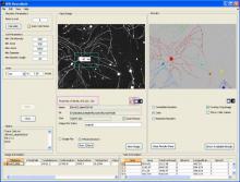
WIS-NeuroMath - is a software tool for automated analysis and quantification of fluorescent microscopy images of Nerve cells, in both in vivo and in vitro preparations. It allows for accurate detection of neurites in challenging images. Following neurite detection, different types of processing can be carried: Cell Morphology of cultured neurons, Neurite Length Analysis and Ganglion Explant Analysis. Usefull also for angiogenesis analysis.

has function
Description
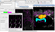
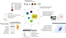
Plugin designed to allow easy semi-automatic tracing of neurons or other filament-like structures (e.g., microtubules, blood vessels) through either 2D images or 3D image stacks. Data can be imported and exported in SWC files for interaction with other software, or details of the traces can be exported as CSV files for analysis in spreadsheets or statistical software. This plugin is included in Fiji by default. A usage instruction is available here: [Tracing Neurons using FIJI (ImageJ)](http://snyderlab.com/2016/05/25/tracing-neurons-using-fiji-imagej/)