Filament tracing

Filament tracing operations are image analysis operations in which there is an image of a filamentous structure (it may be a tree-like structure, a filament network or a agglomeration of single 'stick-like' filaments) as input and outputs data that represent the filament, most commonly a skeleton representation of the filaments and their diameters or surfaces.

Synonyms
Tubular structure extraction
biofilament tracing
Curvilinear structure reconstruction
Curvilinear structure detection
neuron image analysis
neuron reconstruction
Description

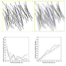
Evaluates the orientation of fiber orientation pattern and plots the results in the image. It calculates gradient in x and y direction. - then calculates the eigenvector of nematic tensor, which is the orientation of the pattern.

Description

Task

Quantify the length of microtubules (MT) and the MT average density per cell.

Workflow descriptions

Simple two step workflow, allowing visual & manual correction of microtubule between the 2 steps. Batch measurement of microtubule lengths for multiple images is achieved by segmenting the MTs and then their skeletonizations. The number of pixels in the microtubule is proportional to their length, so the length can be estimated.

Script

Workflow is written as an ImageJ macro (Fiji) with following steps:

1. The enhancement of tubular structure by computing eigenvalues of the hessian matrix on a Gaussian filtered version of the image ( sigma 1 pixel), as implemented in the tubeness plugin.

2. The tubules were then thresholded , and structures containing less than 3 pixels were discarded.

3. If needed, a visual check and correction of segmented microtubule is then performed.

4. After correction, segmented MTs were then reduced to a 1-pixel thick line using the skeletonize plugin of Fiji. The length of the skeletonized microtubules was then directly proportional to their length.

5. Data were grouped by condition and converted back to micrometers units under Matlab for the statistical tests.

Pitfalls

Commented but not that general without editing some fields in the macros.

Sample Data

Sample data and workflow (see above URL) can be accessed by - login: biii - password Biii!

Misc

3D version also available here. Use of components Skeletonize and Tubeness Filter

need a thumbnail
Description

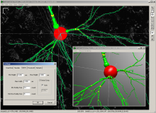
Neuron studio is a software package to reconstruct neurons from 3D confocal images. Reconstruction can be done manually, semi-manually or fully automatic. The images as well as the detected objects are rendered in 3D. A spine detection and classification function is also included. Results can be exported as a text file with coords of the spines. It seems that active development has stopped in 2009. NeuronStudio is being developed at the Computational Neurobiology and Imaging Center (CNIC), a research laboratory at the Neuroscience Department of the Mount Sinai School of Medicine in New York.

NeuronStudio can be used with default parameters or user-defined parameters (Fully or semi-automated).

NeuronStudio_standaloneapp_window_overview
Description

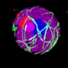
The tool measures the total length of the microtubules in a 3D image.

See: http://dev.mri.cnrs.fr/projects/imagej-macros/wiki/Microtubules_Tool_(3…

You can find a test image here.

3D microtubules