Filament tracing

Filament tracing operations are image analysis operations in which there is an image of a filamentous structure (it may be a tree-like structure, a filament network or a agglomeration of single 'stick-like' filaments) as input and outputs data that represent the filament, most commonly a skeleton representation of the filaments and their diameters or surfaces.

Synonyms
Tubular structure extraction
biofilament tracing
Curvilinear structure reconstruction
Curvilinear structure detection
neuron image analysis
neuron reconstruction
Description

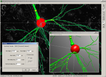
Neuron studio is a software package to reconstruct neurons from 3D confocal images. Reconstruction can be done manually, semi-manually or fully automatic. The images as well as the detected objects are rendered in 3D. A spine detection and classification function is also included. Results can be exported as a text file with coords of the spines. It seems that active development has stopped in 2009. NeuronStudio is being developed at the Computational Neurobiology and Imaging Center (CNIC), a research laboratory at the Neuroscience Department of the Mount Sinai School of Medicine in New York.

NeuronStudio can be used with default parameters or user-defined parameters (Fully or semi-automated).

NeuronStudio_standaloneapp_window_overview
Description

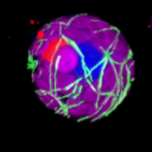
The tool measures the total length of the microtubules in a 3D image.

See: http://dev.mri.cnrs.fr/projects/imagej-macros/wiki/Microtubules_Tool_(3…

You can find a test image here.

3D microtubules
Description

Deprecated component -- superseded by SNT. Legacy description follows:

---

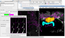
Plugin designed to allow easy semi-automatic tracing of neurons or other filament-like structures (e.g., microtubules, blood vessels) through either 2D images or 3D image stacks. Data can be imported and exported in SWC files for interaction with other software, or details of the traces can be exported as CSV files for analysis in spreadsheets or statistical software.

This plugin is included in Fiji by default.

Description

The Sholl technique is used to describe neuronal arbors. This plugin can perform Sholl directly on 2D and 3D grayscale images of isolated neurons. Its internal algorithm to collect data is based upon how Sholl analysis is done by hand — it creates a series of concentric shells (circles or spheres) around the focus of a neuronal arbor, and counts how many times connected voxels defining the arbor intersect the sampling shells. The major advantages of this plugin over other implementations are:

sholl analysis