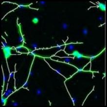
Neuron image analysis

Description

"The plugin analyzes fluorescence microscopy images of neurites and nuclei of dissociated cultured neurons. Given user-defined thresholds, the plugin counts neuronal nuclei, and traces and measures neurite length."[...]" NeuriteTracer is a fast simple-to-use ImageJ plugin for the analysis of outgrowth in two-dimensional fluorescence microscopy images of neuronal cultures. The plugin performed well on images from three different types of neurons with distinct morphologies."

This plugin requires parameter setting: Threshold levels and scale (see more details on the related publication)

Description

We have developed a novel approach, named DF-Tracing, to tackle this challenge. This method first extracts the neurite signal (foreground) from a noisy image by using anisotropic filtering and automated thresholding. Then, DF-Tracing executes a coupled distance-field (DF) algorithm on the extracted foreground neurite signal and reconstructs the neuron morphology automatically. Two distance-transform based “force” fields are used: one for “pressure”, which is the distance transform field of foreground pixels (voxels) to the background, and another for “thrust”, which is the distance transform field of the foreground pixels to an automatically determined seed point. The coupling of these two force fields can“push” a “rolling ball” quickly along the skeleton of a neuron, reconstructing the 3D cell morphology.

Simple Tracing - DT-fields
Description

nctuTW is a "high-throughput computer method of reconstructing the neuronal structure of the fruit fly brain. The design philosophy of the proposed method differs from those of previous methods. We propose first to compute the 2D skeletons of a neuron in each slice of the image stack. The 3D neuronal structure is then constructed from the 2D skeletons. Biologists tend to use confocal microscopes for optimal images in a slice for human visualization; and images in two consecutive slices contain overlapped information. Consequently, a spherical object becomes oval in the image stack; that is, neurons in the image stack do not reflect the true shape of the neuron. This is the main reason we chose not to work directly on the 3D volume.

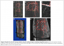
The proposed method comprises two steps. The first is the image processing step, which involves computing a set of voxels that is a superset of the 3D centerlines of the neuron. The shortest path graph algorithm then computes the centerlines. The proposed method was applied to process more than 16 000 neurons. By using a large amount of reconstructions, this study also demonstrated a result derived from the reconstructed data using the clustering technique." (Extracted from reference publication)

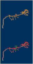
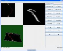
Illustrative image shows gold standard (top) and method results (bottom). 

nctuTW_results_example
Description

By combining multiple image alignment and tracing into one program, Reconstruct (TM) allows images to be processed more efficiently. Tracing can be done directly on the transformed images and alignments can be asily modified. Reconstruct (TM) was developed from years of experience working with high magnification serial section images of brain tissue. (Extracted from User Manual)

"The original platform of the Reconstruct program allows a user to trace objects in serial sections by manually drawing the outline of each object on each section, which is time-consuming. We modified Reconstruct to enable semi-automatic tracing of axons using a region-growing algorithm called wildfire."

Reconstruct_standaloneapp_example_Results
Description

JFilament is an ImageJ plugin for segmentation and tracking of 2D and 3D filaments in fluorescenece microscopy images. The main algorithm used in Jfilament is "Stretching Open Active Contours" (SOAC). In order to use this method, the user must define seed points in the image where the SOAC method will begin.

JFilament also includes 2D "closed" active contours which can be used for tasks such as segmentation and tracking of cell boundaries.


JFilament_ImageJ_pulgin_Window