Light-sheet microscopy

Synonyms
LSFM
Digitally scanned Lightsheet Microscopy
Selective Plane Illumination Microscopy
Light-sheet fluorescence microscopy
DSLM
SPIM
Lattice Light-sheet Microscopy
Dual-View inverted SPIM
Spherical aberrations assisted Extended Depth-of-field Lightsheet Microscopy
Bessel Beam Lightsheet Microscopy
single objective Selective Plane Illumination Microscopy
Hardware implementations: multidirectional SPIM
LLSM
Clarity Optimized Lightsheet Microscopy
Multiview Selective Plane Illumination Microscopy
MuViSPIM
inverted SPIM
soSPIM
diSPIM
COLM
SPED
mSPIM
iSPIM
Description

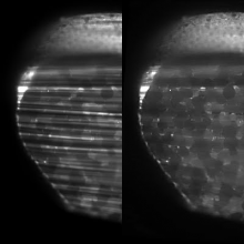
This macro implements a filter that is meant to attenuate close to parallel intensity stripes in an image, such as often happening in light sheet microscopy. The results are usually decent even when the stripes show a large angular spread due to light sheet refraction at the sample surface. The filter can process a 3D stack but the processing is performed slice by slice.

Example image is available in the documentation link. 

Description

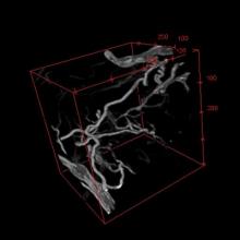
This macro segments blood vessels in a 3D stack. It is suited for well-contrasted images (low background) and works better if the width of the vessels of interest is reasonably uniform.


Sample image: 1

sample image: 2

has function
Description

The Huygens Software Suite consists of different image processing packages with functionalities that include deconvolution, interactive analysis, and volume visualization of 2D-3D multi-channel and time series images from fluorescence microscopes such as widefield, confocal, multi-photon, spinning disk, Array Detector, STED, and Light Sheet

Description

**Collaborative Annotation Toolkit for Massive Amounts of Image Data** CATMAID is a Collaborative Annotation Toolkit for Massive Amounts of Image Data. It is designed to navigate, share and collaboratively annotate massive image data sets of biological specimens. The interface is inspired by GoogleMaps, with which it shares basic navigation concepts, enhanced to allow the exploration of 3D biological image data acquired by optical or physical sectioning microscopy techniques. The interface enables seamless sharing of regions of interest through bookmarks and synchronized navigation through multiple registered data sets. With massive biological image data sets it is unrealistic to create a sustainable centralized repository. A unique feature of CATMAID is its partially decentralized architecture where the presented image data can reside on any Internet accessible server and yet can be easily cross-referenced in the central database. In this way no image data are duplicated and the data producers retain full control over their images. CATMAID is intended to serve as data sharing platform for biologists using high-resolution imaging techniques to probe large specimens. Any high-throughput, high-content imaging project such as gene expression pattern screens would benefit from the interface for data sharing and annotation.

CATMAID
Description

This plugin combines the idea of using fiduciary markers, local descriptors and geometric hashing and applies global optimization. It can register an arbitrary number of partially overlapping point clouds. It is robust with respect to the amount of incorporated beads, bead distribution, amount of overlap, and can reliably detect non-affine disturbances (e.g. abrupt agarose movement) that might occur during imaging.

For details about the SPIM registration, fusion & deconvolution please have a look at the Multiview Reconstruction Plugin. It is much more powerful, flexible and completely integrated with the BigDataViewer.

need a thumbnail