Focal Adhesion

Description

Junction Mapper is a semi-automated software (Java Desktop application) for analysing data from images of cells in close proximity to each other in monolayers. The focus of Junction Mapper is to measure the morphology of cell boundaries, define single junctions and quantify the length, area and intensity of the staining of different proteins localised at cell-cell contacts. The output are various unique parameters that assess the contacting interface between cells and up to two junctional markers.

junction mapper
Description

The website implements a set of computer vision algorithms designed to automatically process time-lapse images of fluorescently labeled focal adhesion proteins in motile cells.

The methods associated with the processing have been published in PLOS One and Cell. The publication describes a quantitative analysis of focal adhesion dynamics that have been imaged using TIRF. All image processing steps are well explained or referenced.

To better understand the dynamic regulation of focal adhesions, we have developed an analysis system for the automated detection, tracking, and data extraction of these structures in living cells. This analysis system was used to quantify the dynamics of fluorescently tagged Paxillin and FAK in NIH 3T3 fibroblasts followed via Total Internal Reflection Fluorescence Microscopy (TIRF). High content time series included the size, shape, intensity, and position of every adhesion present in a living cell. These properties were followed over time, revealing adhesion lifetime and turnover rates, and segregation of properties into distinct zones.


has function
Description

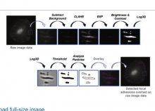
Tracking of focal adhesions includes a number of challenges:

  1. Detection of focal adhesion regions in areas of highly variable background
  2. Separation of "clumped" adhesions in different objects.
  3. Dynamics: Focal adhesions dynamically, grow, shrink, change their shape, they can fuse with neighboring adhesions or one adhesion can be split into multiple children.

Würflinger et al (2011) describe how to detect focal adhesion objects and how to track them over time. Interestingly, tracking results are fed back to segmentation to improve separation of clumped adhesions.

The authors implemented the workflow in Matlab, but do not provide a ready-to-use script.

Description

The quantification is explained in detail in chapter 8 "Cell Polarity - Focal Adhesion and Actin Dynamics in Migrating Cells" in "Bioimage Data Analysis Book" downloadable from here.

For codes and sample images, download the zipped archive (linked under "Download").

has function
Description

Simple workflow description for ImageJ, step-by-step description for delineating focal adhesions, count and characterize their positions.  

Measurement of dynamics is not involved.