multi-channel

Description

This macro can stitch a (Z,T,C) data set with virtually no limit on the number of Z slices and time frames. The input to the macro is a folder with the raw tiff images (one image per file) as typically exported by motorized microscopes. These files must all be stores in the same folder and the file naming should ideally comply to OME-TIFF. The macro is however quite flexible: Only --X, --Y and --Z fields with user defined number of digits are compulsory. --T, --C and --L fields with user defined number of digits are necessary for multiple time frames / channels data sets. A compatible data set is provided as a .zip archive. Before processing it unzip it to a given location. The stitching is performed in a reference Z slice (and in a specific reference time frame and channel). The same displacements are applied to all the Z slices, time frames and channels. Before starting the batch processing a montage with the original images of the selected Z slice / time frame / channel is displayed together with the stitched image in this stack. If you are not satisfied with the result you can select another reference. The stitching is then performed time frame by time frame and slice by slice and the stitched images are exported to a single user defined output folder. The macro can also process a data set with multiple channels, the stitching is then computed once on a reference channel and then applied to the other channels.

has topic
has function
Description

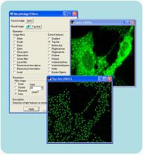
WASH, Exo84, and cortactin spot detection and codistribution analysis To detect endosomes, an automatic Otsu threshold is applied to the Gaussian-filtered MT1-MMP–positive endosome image (= 1.5 pixels for the sample image). Statistics about each endosome are then saved, for example random positioning of spots can be compared to actual positioning. For each endosome, WASH and Exo84 (or WASH and cortactin) spots are searched for in a neighboring of x pixels in their respective channel. Their number and position are saved per endosome (**see the macro in Text file S2 downloadable from here**).

From the position of WASH and Exo84 (or WASH and cortactin) spots around each endosomes, each WASH spot is paired with its closest Exo84 (or cortactin) spot neighbor, optimized over all spots around this endosome.

This allowed measuring of the distribution of distance between WASH-Exo84 (or WASH-cortactin) spots (**for the co-distribution analysis, see matlab scripts in Zip file S3 downloadable).

endosomes and spot neighbors
Description

ITK-SNAP is a software application used to segment structures in 3D medical images. It can also be used as a 3D annotation tool for deep learning. It is based on ITK, VTK libraries.

Description

Metamorph provides all tools needed to perform analysis of acquired images with user-friendly application modules for biology-specific analysis such as cell signaling, cell counting, and protein expression.

Matamorph user interface
Description

The Huygens Software Suite consists of different image processing packages with functionalities that include deconvolution, interactive analysis, and volume visualization of 2D-3D multi-channel and time series images from fluorescence microscopes such as widefield, confocal, multi-photon, spinning disk, Array Detector, STED, and Light Sheet