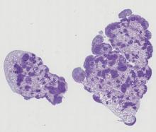
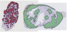
tissue

Human colon tissue

Submitted by Perrine on Mon, 08/26/2019 - 11:30

One of the principal challenges in counting or segmenting cells or cell nuclei is dealing with clustered objects such as in tissues. To help assess algorithms' performance in this regard, synthetic 3D image sets of human colon tissue are provided in two diferent levels of quality: high SNR and low SNR. Ground truth is available as well.

Description

Orbit Image Analysis is a free open source software with the focus to quantify big images like whole slide scans.

It can connect to image servers, e.g. Omero.
Analysis can be done on your local computer or via scaleout functionality in a distrubuted computing environment like a Spark cluster.

Sophisticated image analysis algorithms incl. tissue quantification using machine learning, object segmentation and classification are build in. In addition a versatile API allows you to enhance Orbit and to run your own scripts.

Orbit
Description

QuantCenter is the framework for 3DHISTECH image analysis applications. with the goal of helping the pathologists to diagnose in an easier way. QuantCenter, is optimized for whole slide quantification. It has a linkable algorithm concept that tries to provide an easy-to-use and logical workflow. The user has different quantification modules that he or she could link one after other to fine-tune or to speed up the analysis.

QuantCenter logo
Description

This is the "prediction step" of the Pyxit segmentation model builder. It is a learnable segmentation algorithm based on ground-truth images and segmentation mask. It learns a multiple output pixel classification algorithm. It downloads from Cytomine-Core annotation images+alphamasks from project(s), build a segmentation (pixel classifier) model which is saved locally. Typical application: tumor detection in tissues in histology slides. 

Pyxit example
Description

This is a learnable segmentation algorithm based on ground-truth images and segmentation mask. It learns a multiple output pixel classification algorithm. It downloads from Cytomine-Core annotation images+alphamasks from project(s), build a segmentation (pixel classifier) model which is saved locally. Typical application: tumor detection in tissues in histology slides. It is based on "Fast Multi-Class Image Annotation with Random Subwindows and Multiple Output Randomized Trees" http://orbi.ulg.ac.be/handle/2268/12205 and was used in "A hybrid human-computer approach for large-scale image-based measurements using web services and machine learning" http://orbi.ulg.ac.be/handle/2268/162084?locale=en

Segmentation illustration