Free and open source

Description
Python-bioformats is a Python wrapper for Bio-Formats, a standalone Java library for reading and writing life sciences image file formats. Bio-Formats is capable of parsing both pixels and metadata for a large number of formats, as well as writing to several formats. Python-bioformats uses the python-javabridge to start a Java virtual machine from Python and interact with it. Python-bioformats was developed for and is used by the cell image analysis software CellProfiler (cellprofiler.org). PyPI record: https://pypi.python.org/pypi/python-bioformats Documentation: http://pythonhosted.org/python-bioformats/ GitHub repository: https://github.com/CellProfiler/python-bioformats Report bugs here: https://github.com/CellProfiler/python-bioformats/issues python-bioformats is licensed under the GPL license to be compatible with the copy of Bio-Formats that is distributed with the package, but is compatible with a BSD license if loci_tools.jar is replaced with SCIFIO jars. See the accompanying file LICENSE for details.
need a thumbnail
Description

## Features >The IJBlob library indentifying connected components in binary images. The algorithm used for connected component labeling is: >Chang, F. (2004). A linear-time component-labeling algorithm using contour tracing technique. Computer Vision and Image Understanding, 93(2), 206–220. doi:10.1016/j.cviu.2003.09.002 ##Reference Wagner, T and Lipinski, H 2013. IJBlob: An ImageJ Library for Connected Component Analysis and Shape Analysis. Journal of Open Research Software 1(1):e6, DOI:

need a thumbnail
Description

A workflow combining ImageJ macro and manually using Trainable Weka Segmentation plugin for counting clumped cells.

Description

An ImageJ macro for calculating empty surfaces on histological slices (ex: tubules in a kidney).

has topic
Description
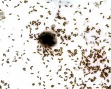
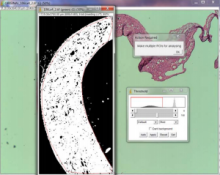
<p>Particle detection is based on "Analyze Particles" in ImageJ. It probably could also be used in spot detection, not limited to centromere. &gt;This macro is described in Bodor et al. (2012). The macro recognizes centromere or kinetochore foci in Delta Vision or TIFF images and determines their centroid position. Fluorescent intensities are then measured for each centromere by placing a small box around the centroid position of the centromere. The peak intensity value within the box is corrected for local background by subtraction of the minimum pixel value. This process results in an accurate measurement of large numbers of centromere or kinetochore-specific signals. Following papers uses CRaQ (picked up, maybe more): - [Fachinetti et al. (2017)](https://www.cell.com/developmental-cell/pdf/S1534-5807(16)30909-1.pdf), Developmental Cell 40, 104–113, - [Guo et al. (2017)](https://www.nature.com/articles/ncomms15775) Nature Communications volume 8, Article number: 15775 (2017) doi:10.1038/ncomms15775 - [Lgosdon et. al. (2015)](http://jcb.rupress.org/content/208/5/521) J Cell Biol Mar 2015, 208 (5) 521-531; DOI: 10.1083/jcb.201412011 - [Bodor et al. (2014)](https://www.ncbi.nlm.nih.gov/pmc/articles/PMC4091408/), eLife. 2014; 3: e02137</p>
has function