multi-channel

Description

This ImageJ plug-in is a compilation of co-localization tools. It allows:

-Calculating a set of commonly used co-localization indicators:

Pearson's coefficient Overlap coefficient k1 & k2 coefficients Manders' coefficient Generating commonly used visualizations:

-Cytofluorogram

Having access to more recently published methods:

-Costes' automatic threshold

Li's ICA Costes' randomization Objects based methods (2 methods: distances between centres and centre-particle coincidence)

example of partial colocalisation from reference publication
Description

Bio Image Analysis tool from REF

logo ImageJ
Description

CellProfiler is free, open-source software for quantitative analysis of biological images.

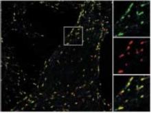
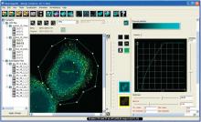
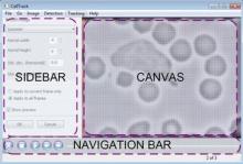
CellProfiler is designed to enable biologists without training in computer vision or programming to quantitatively measure cell or whole-organism phenotypes from thousands of images automatically. The researcher creates an analysis pipeline from modules that find cells and cell compartments, measure features of those cells to form a rich, quantitative dataset that characterizes the imaged site in all of its heterogeneity. CellProfiler is structured so that the most general and successful methods and strategies are the ones that are automatically suggested, but the user can override these defaults and pull from many of the basic algorithms and techniques of image analysis to solve harder problems. CellProfiler is extensible through plugins written in Python or for ImageJ. Strengths: Cells, Neurons, C. Elegans, 2D Fluorescent images, high-throughput screening, phenotype classification, multiple stains/site, interoperability, extensibility, machine learning, segmentation Limitations: largely limited to 2D, not well suited to manually-guided analysis or content review, image size limitations

Description

BioImageXD is a free open source software package for analyzing, processing and visualizing multi-dimensional microscopy images. It's a collaborative project, designed and developed by microscopists, cell biologists and software engineers from the Universities of Jyväskylä and Turku in Finland, Max Planck Institute CBG in Dresden, Germany and collaborators worldwide. BioImageXD was published in the July 2012 issue of Nature Methods.

Screen capture of BioImageXD
Description

A standalone cell tracking software for single cell migration. Tracking of cells in tissue was also done in Drosophila germband.

GUI image (from http://sacan.biomed.drexel.edu/celltrack)