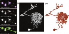
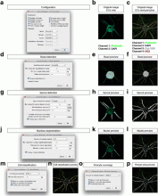
Component

A Component is an implementation of certain image processing / analysis algorithms.

Each component alone does not solve a Bioimage Analysis problem.

These problems can be addressed by combining such components into workflows.

Description

"We have developed an automatic graph algorithm, called the all-path pruning (APP), to trace the 3D structure of a neuron. To avoid potential mis-tracing of some parts of a neuron, an APP first produces an initial over-reconstruction, by tracing the optimal geodesic shortest path from the seed location to every possible destination voxel/pixel location in the image. Since the initial reconstruction contains all the possible paths and thus could contain redundant structural components (SC), we simplify the entire reconstruction without compromising its connectedness by pruning the redundant structural elements, using a new maximal- covering minimal-redundant (MCMR) subgraph algorithm. We show that MCMR has a linear computational complexity and will converge. We examined the performance of our method using challenging 3D neuronal image datasets of model organisms (e.g. fruit fly)"

This plugin can be used with default parameters or user-defined parameters.

APP_Vaa3D_example_results
Description

The Sprout Morphology plugin measures sprout number, length, width and cell density of endothelial cell (EC) sprouts grown in a bead sprouting assay. It optionally includes measuring the coverage of these sprouts with pericytes included in the assay, as well as the endothelial cell/pericyte ratio.

graphical abstract
Description

Spot detector detects and counts spots, based on wavelet transform.

- Detects spots in noisy images 2D/3D.
- Depending on objective, spots can be nuclei, nucleus or cell
- Versatile input: sequence or batch of file.
- Detects spot in specific band/channel.
- Multi band labeling: automaticaly creates ROIs from one band and count in the same or an other band.
- Filters detection by size.
- Sort detection by ROIs
- Output data in XLS Excel files: number of detection by ROIs, and each detection location and size.
- Outputs withness image with ROIs and detection painted on it.
- Outputs binary detection image.
- Displays detections
- Displays tags

logo spot detector
Description

This ImageJ plug-in is a compilation of co-localization tools. It allows:

-Calculating a set of commonly used co-localization indicators:

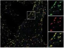
Pearson's coefficient Overlap coefficient k1 & k2 coefficients Manders' coefficient Generating commonly used visualizations:

-Cytofluorogram

Having access to more recently published methods:

-Costes' automatic threshold

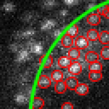
Li's ICA Costes' randomization Objects based methods (2 methods: distances between centres and centre-particle coincidence)

example of partial colocalisation from reference publication
Description

SRRF is a high-performance analytical approach for Live-cell Super-Resolution Microscopy, provided as a fast GPU-enabled ImageJ plugin. SRRF is capable of extracting high-fidelity super-resolution information from TIRF, widefield and confocals using conventional fluorophores such as GFP. SRRF is capable of live-cell imaging over timescales ranging from minutes to hours.

Comparison TIRF - SRRF