Collection

A collection is a software that encapsulate a set of bioimage components and/or workflows.

Description

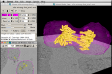
IMOD is a set of image processing, modeling and display programs used for tomographic reconstruction and for 3D reconstruction of EM serial sections and optical sections. The package contains tools for assembling and aligning data within multiple types and sizes of image stacks, viewing 3-D data from any orientation, and modeling and display of the image files.

Included are two programs with graphical interface: 3dmod, for displaying and segmenting 2D images and 3D volumes; etomo, for reconstructing tomographic volumes from tilt series of images.

Processing can be distributed on multiple cores and executed in batch mode.

iMod
Description

MATLAB is famous, so this page is only for being the landing page for components and workflows.

Matlab logo
Description

Acquiarium is for carrying out the common pipeline of many spatial cell studies using fluorescence microscopy. It addresses image capture, raw image correction, image segmentation, quantification of segmented objects and their spatial arrangement, volume rendering, and statistical evaluation. It is focused on quantification of spatial properties of many objects and their mutual spatial relations in a collection of many 3D images. It can be used for analysis of a collection of 2D images or time lapse series of 2D or 3D images as well. It has a modular design and is extensible via plug-ins. It is a stand-alone, easy to install application written in C++ language. The GUI is written using cross-platform wxWidgets library.

Functionalities
Description

Columbus is a combination of an image database (based on Omero, OME) and an image analysis engine based on Acapella (PerkinElmer). It is dedicated to cell culture based high content screening data and is used via a web interface. It provides a set importers for automated microscopes such as Yokogawa CellVoyager, PerkinElmer Operetta, PerkinElmer Opera and data in Metamorph format. After login, Images can be explored in a standard web browser by clicking on a well plate view. Image analysis workflows can be developed by combining modules like "find nuclei", "find cytoplasm", "find spots" for object detection. Objects can have a hierarchical structure, e.g. spot objects can be part of a cell object. The approach of workflow design is similar to the freeware cell profiler, but more restricted (less functions and less parameters to tweak) and easier to use. Mutliple intensity- and shape based features can be calculated from detected objects (e.g. texture: haralick, Garbor, SER). Objects can be classified by these features by using hard thresholds or by supervised machine learning. Analysis workflows and results are stored in the database and can be exprted as csv tables for secondary analysis. Simple secondary analysis workflows can be also applied in Columbus directly. Results can be visualized as heatmaps on the plate view. The HCS statistics software Genedata Screener Assay Analyzer can be directly connected to the database.

Columbus screenshot
Description

Slicer, or 3D Slicer, is a free, open source software package for visualization and image analysis. 3D Slicer is natively designed to be available on multiple platforms, including Windows, Linux and Mac Os X.

3D slicer