2D

Description

This workflow classifies objects based on object-level features (e.g. intensity based, morphology based, etc) and user annotations. It needs segmentation images besides the raw image data. Segmentation images can be obtained from ilastik pixel classification, or binary segmentation images from other tools. Within the object classification, one can prefilter objects through thresholds (on pixel probability image) or object sizes (on segmentation image). Outputs are predicted classification label images. Selected features can also be exported. Advanced users also have possibilities to add customized (object) features for classification in a simple plugin fashion through python scripts.

Description

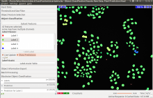
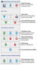
This workflow is used to track multiple (appear/disappear, dividing and merging) objects in presumably big 2D+t or 3D+t datasets. It is best suitable for roundish objects or spots. Tracking is done through segmentation, which can be obtained from ilastik pixel classification, or imported from other tools. Users should provide a few object level labels, and the software predicts results on the rest of the image or new images with similar image characteristics. As a result, all objects get assigned random IDs at the first frame of the image sequence and all descendants in the same track (also children objects such as daughter cells) inherit this ID.

need a thumbnail
Description

The Artemia Tools help to calculate the normalized redness of Artemia in color images.

See: http://dev.mri.cnrs.fr/projects/imagej-macros/wiki/Artemia_Tools

Test images: http://biii.eu/node/1139

Artemia color analysis toolset
Description

This tool allows for extraction of image series from Olympus Slide Scanners. These VSI files usually contain several images that are too big to load into memory (>50k x 50k pixels). It was written and tested on Fiji and is available from a Fiji Update Site: http://fiji.sc/List_of_update_sites

has function
VSI screenshot
Description

In this human cytoplasm-nucleus translocation assay, learn how to load a previously calculated illumination correction function for two separate channels, measure protein content in the nucleus and cytoplasm, and calculate the ratio as a measure of translocation. This is a clumpy cell type, so studying the settings in primary object identification may be helpful for users interested in the more advanced options that module offers. More about these images can be found at the BBBC.

need a thumbnail