Component

A Component is an implementation of certain image processing / analysis algorithms.

Each component alone does not solve a Bioimage Analysis problem.

These problems can be addressed by combining such components into workflows.

Description

This module is for learning classification models from ground-truth data (supervised learning). It downloads from Cytomine-Core annotation images and coordinate of annotated objects from project(s) and build a annotation classification model which is saved locally.  

It is used by Cytomine DataMining applications: classification_validation, classification_model_builder, classification_prediction, segmentation_model_builder and segmentation_prediction. But it can be run without Cytomine on local data (using dir_ls and dir_ts arguments).

has topic
need a thumbnail
Description

Automatic registration in 2D or 3D based on detection or binary mask. Takes images with detections already done on it.

logo autofinder
Description

This plugin allows to compute a similarity (translation/rotation/scaling and flipping) transform from pair of points. It is updating the transformed image interactively such that the user get immediate feedback. The transformation is saved and can be applied to any other stack/image. Non rigid deformation can also be applied in 2D or 3D.

3D/3D,2D/3D or 3D /2D can be handled .

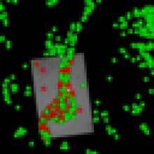
3D ROI are enabled, and can be checked with the 3D vtk view (size of ROI can be changed using the ROI stroke width).

Some prealignment by rotating in 3D the volume is possible.

Transformations can be applied directly or combined through Block Protocols (search for apply transformation).

It's also provide information about the predicted Error (based on statistical prediction), either as a full color mapping, either on each points used as landmarks, and error on the discrepancy in position between points.

There are video tutorials available in the web.


logo ec-clem
Description

A resident function in ImageJ, located in the menu as [Process > Binary > Voronoi].

Quote from the ImageJ reference page:

Splits the image by lines of points having equal distance to the borders of the two nearest particles. Thus, the Voronoi cell of each particle includes all points that are nearer to this particle than any other particle. When particles are single points, this process is a Voronoi tessellation (also known as Dirichlet tessellation). The output type (Overwrite, 8-bit, 16-bit or 32-bit) of this command can be set in the [Process > Binary > Options...] dialog box. In the output, the value inside the Voronoi cells is zero; the pixel values of the dividing lines between the cells are equal to the distance between the two nearest particles. This is similar to a medial axis transform of the background, but there are no lines in inner holes of particles.