CellProfiler Pipeline

Description

In this example, cells are grown as a tissue monolayer. Rather than identifying individual cells, this pipeline quantifies the area occupied by the tissue sample.


Download package also contains example images. 

Description

quote

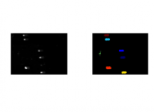
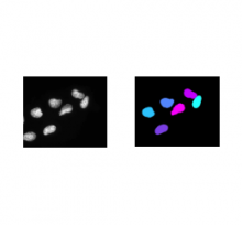
This is a simple example of a DNA damage assay using single cell gel electrophoresis. Here, the measurement of interest is the length and intensity of the comet tail. Also, illumination correction is used to reduce background fluorescence prior to measurement. Also shown is a silver-stained comet example in which the percentage of DNA contained in the tail is calculated.

Example Images: Packaged together with the cellprofiler pipeline file. 

Description

The goal of this workflow is to track cells captured in a time-lapse movie of a syncytial blastoderm stage Drosophila embryo and quantify their movement.

This example shows an example of object tracking. This pipeline analyzes a time-lapse experiment to identify the cells and track them from frame to frame, which is challenging since the cells are also moving. In addition, this pipeline also extracts metadata from the filename and uses groups the images by metadata in order to independently process several sequences of images and output the measurements of each.

Sample images

A portion of a time lapse movie of a syncytial blastoderm stage Drosophila embryo with a GFP-histone gene which renders chromatin fluorescent in live embryos. The movie shows nuclear divisions 10 through 13.

Victoria Foe made this movie on a Bio-Rad Radiance 2000 laser scanning confocal microscope using a 40X 1.3NA oil objective. The frames are 7 seconds apart and plays at 30 frames per second

GFP-histone transformed files provided by Rob Saint

V.Foe and G.Odell, . 26 July 2001

has function
Description

Quote:

This pipeline shows how to identify smaller objects (foci) within larger objects (nuclei) and how to use the Relate module to establish a relationship between the two as well as perform per-object aggregate measurements (such as number of foci per nucleus). Sample images are included in the download package.

Description

Quote:

Measuring the colocalization between fluorescently labeled molecules is a widely used approach to measure the degree of spatial coincidence and potential interactions among subcellular species (e.g., proteins). This example shows how the object identification and RelateObjects modules are used to measure the degree of overlap between two fluorescent channels. Sample image is included in the download package.