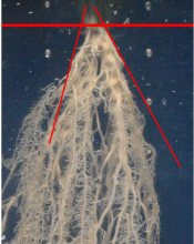
ImageJ Macros

Description

Count bacterial colonies on agar plates and measure the occupied surfaces. The user has to provide a selection (roi) of the area that will be analyzed. He can than run the segmentation and if necessary correct the results. In a third step he can run the counting and measurement.

has function
Description

Normalize the orientation of the images of the Zebrafish embryos.

In the documentation webpage, the aim of the workflow is to normalize the orientation of the images of the Zebrafish embryos, find the point of injection of tumor cells and measure the distribution of Cy3 stained tumor foci.

ImageJ macro implementation of the Workflow described in Ghotra et al (2012). Note that currently only the angle and orientation normalization is implemented in this version.

Sample images are linked in the documentation webpage. 

has function
Description

The workflow measures the growth of cells in 3D, combining an ImageJ macro for preprocessing and successive tracking using Imaris.  

The sample dataset (available in the github repository) contains 2-Photon images of neurons. The neurons were imaged in 3D at two time frames.To allow measuring significant differences in cell volume, the time gap between the frames is large (ca. 30 min) and the animal was removed in the waiting phase. For this reason, there is a considerable shift in sample position between the frames that has to be corrected before cell detection and tracking.

The workflow consists of following steps:

1. Import of single tiff slices [imageJ macro]

2. Organizing the data in a 4D time series with 2 time frames [imageJ macro]

3. Correction of shift between the time frames by rigid registration [imagJ macro]

4. Bleaching correction [imageJ macro]

5. Export of preprocessed image data in ics/ids format [imageJ macro]

6. Import of ics/ids data to Imaris [Imaris]

7. Cell object detection as "Imaris Surface Object" [Imaris]

8. Tracking cell objects over time [Imaris]

9. Split Tracks (use Imaris XT extension "Split Tracks") to generate single cell objects [Imaris]

10. Export the statistics: Select the complete folder, go to the statistics tab and use ‚Full Export’ [Imaris]

The preprocessing macro can be referenced here.

The sample images were acquired by Cordula Ulbrich (Petzold Group at German Center of Neurodegenerative Disesases (DZNE), Bonn, Germany).

Input data type: tiff

Output data type: data table

has function
Description

The linked webpage presents a collection of ImageJ macros for Intelligent Imaging (Feedback to microscope system for the secondary scan). 

An ImageJ macro able to control some microscopes (Micro-manager or Leica CAM controlled) to acquire high resolution images of only some structures (e.g. isolated cells) or events (e.g. mitosis) within a sample. The scan is sequenced as a primary (low resolution monitoring) scan and a secondary (high resolution, multi-dimensional) scan.

has function
Description

This macro is a plugin macro to the "Intelligent Imaging" workflow. It detects the Cytoo patterns (specific fluorsecence channel) and computes the occupancy (number of cells) of each pattern by analyzing the images of the DAPI channel. The analysis function can be easily extended to, for instance, only select the cells that are well spread on the patterns (by analyzing a third channel with a properly chosen marker of the cytoplasm).

need a thumbnail