Workflow

A workflow is a set of components assembled in some specific order to

  1. Measure and estimate some numerical parameters of the biological system or
  2. Visualization

for addressing a biological question. Workflows can be a combination of components from the same or different software packages using several scripts and manual steps.

Description

"The plugin analyzes fluorescence microscopy images of neurites and nuclei of dissociated cultured neurons. Given user-defined thresholds, the plugin counts neuronal nuclei, and traces and measures neurite length."[...]" NeuriteTracer is a fast simple-to-use ImageJ plugin for the analysis of outgrowth in two-dimensional fluorescence microscopy images of neuronal cultures. The plugin performed well on images from three different types of neurons with distinct morphologies."

This plugin requires parameter setting: Threshold levels and scale (see more details on the related publication)

Description

Integrates hardware control of Leica microscopes (via CAM), image analysis (e.g. via ImageJ, Matlab), and adaptive automatic screening of identified regions of interest.

Description

The wound healing tool measures the area of a wound in a time series of images of cellular tissue. The tool will measure the area of the wound, i.e. the area that does not contain tissue, in each image. The segmentation is based on the fact that the image is more homogeneous in the region of the wound as in the region of the tissue. Via the options, one of two methods to detect the empty area, can be selected. The first uses edge detection, the second a variance filter. Holes in the detected tissue are filled using morphological operations.

Measure area of the wound
Description

The skin tools measure the thickness of the epidermis and the interdigitation index.

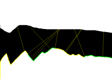
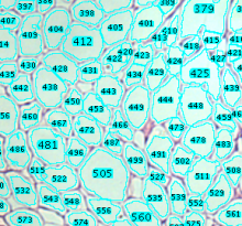
The input images are masks that represent the epidermis and that have been created from images of stained histological sections. The mask must touch the left and right border of the image. The dermal-epidermal border must be on the lower site of the image. The interdigitation index can be measured for one or more segments per image. As a measure of the thickness of the epidermis the lengths of a number of random line segments are measured. The line segments start at the lower border, are perpendicular to the lower border and end at the opposite border of the mask.

See installation Instructions on the website.

has topic
Measure thickness from a mask
Description

The Adipocytes Tools help to analyze fat cells in images from histological section. This is a rather general cell segmentation approach. It can be adapted to different situations via the parameters. This means that you have to find the right parameters for your application.

Sample Image: [0178_x5_3.tif](http://dev.mri.cnrs.fr/attachments/190/0178_x5_3.tif)

has topic
has function