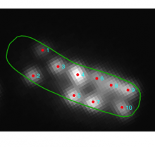
Oufti (previously named MicrobeTracker) is a MATLAB application / suite of tools for analysing fluorescent spots inside microbes. MicrobeTracker can identify cell outlines and fluorescent foci, and generate plots and statistics based on positions and intensity (kymographs, histograms etc.) The MATLAB code is easy to modify and extend to add additional plots and statistics: see e.g. Lesterlin et al. (2014).
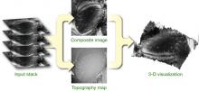
Wavelet-based method to merge a stack of micrographs taken at different focal positions (aligned along the optical axis) into a single, entirely focused composite image.
This plugin provides an extended depth of field algorithm to obtain in focus microscopic images of 3D objects and organisms using different algorithms: Sobel, variance, real and complex wavelets.
This plugin is bundled with Fiji. For installation in ImageJ1, download from the link below and manually install the class file.
Quote:
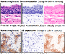
The colour deconvolution plugin (java and class files) for ImageJ and Fiji implements stain separation using Ruifrok and Johnston's method described in [1]. The code is based on a NIH Image macro kindly provided by A.C. Ruifrok.
The plugin assumes images generated by colour subtraction (i.e. light-absorbing dyes such as those used in bright field histology or ink on printed paper). However, the dyes should not be neutral grey (most histological stains are not so).
If you intend to work with this plugin, it is important to read the original paper to understand how new vectors are determined and how the procedure works.
The plugin works correctly when the background is neutral (white to grey), so background subtraction with colour correction must be applied to the images before processing.
The plugin provides a number of "built in" stain vectors some of which were determined experimentally in our lab (marked in the source with GL), but you should determine your own vectors to achieve an accurate stain separation, depending on the stains and methods you use. See the note below.
The built-in vectors are :
Haematoxylin and Eosin (H&E) x2
Haematoxylin and DAB (H DAB)
Feulgen Light Green
Giemsa
Fast Red, Fast Blue and DAB
Methyl green and DAB
Haematoxylin, Eosin and DAB (H&E DAB)
Haematoxylin and AEC (H AEC)
Azan-Mallory
Masson Trichrome
Alcian blue & Haematoxylin
Haematoxylin and Periodic Acid - Schiff (PAS)
RGB subtractive
CMY subtractive
User values entered by hand
Values interactively determined from rectangular ROIs