3D

Description

This macro extracts .lei and .lif multichannel Z-stacks into multiple .tif stacks, splitting the channels into different stacks. Several options are possible such as background substraction, various filters, or optional reset of spatial scale. Requires Bio-Formats plugin

Description

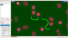
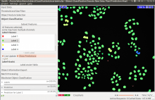
This workflow classifies, or segments, the pixels of an image given user annotations. It is especially suited if the objects of interests are visually (brightness, color, texture) distinct from their surrounding. Users can iteratively select pixel features and provide pixel annotations through a live visualization of selected feature values and current prediction responses. Upon users' satisfaction, the workflow then predicts the remaining unprocessed image(s) regions or new images (as batch processing). Users can export (as images of various formats): selected features, annotations, predicted classification probability, simple segmentation, etc. This workflow is often served as one of the first step options for other workflows offered by ilastik, such as object classification, automatic tracking.

Description

This workflow classifies objects based on object-level features (e.g. intensity based, morphology based, etc) and user annotations. It needs segmentation images besides the raw image data. Segmentation images can be obtained from ilastik pixel classification, or binary segmentation images from other tools. Within the object classification, one can prefilter objects through thresholds (on pixel probability image) or object sizes (on segmentation image). Outputs are predicted classification label images. Selected features can also be exported. Advanced users also have possibilities to add customized (object) features for classification in a simple plugin fashion through python scripts.

Description

This workflow is used to track multiple (appear/disappear, dividing and merging) objects in presumably big 2D+t or 3D+t datasets. It is best suitable for roundish objects or spots. Tracking is done through segmentation, which can be obtained from ilastik pixel classification, or imported from other tools. Users should provide a few object level labels, and the software predicts results on the rest of the image or new images with similar image characteristics. As a result, all objects get assigned random IDs at the first frame of the image sequence and all descendants in the same track (also children objects such as daughter cells) inherit this ID.

need a thumbnail
Description

A simple workflow is described in the following for measuring subcellular localizations of organelle by the distance from the nucleus. For example, you can quantify how far some type of vesicles or protein aggregates are apart from the nucleus border. This workflow is for analyzing 3D data.

Data requirements:

  • 3D data, 2 channels
  • Channel 1: nucleus stain = Channel 2: stain for marker you want to quantify the distance to nucleus for

Workflow:

  1. Nucleus detection: Imaris
  • Add a new SURFACE object, name it "nuclei"
  • Follow the object detection wizard to segment nucleus objects
  1. Marker object detection: Imaris
  • Add a new SURFACE object
  • Follow the object detection wizard to segment nucleus objects
  1. Creating of distance map channel: Imaris
  • In the image processing menu, go to SurfacesFunctions>>Distance Transformation
  1. MATLAB:
  • select nucleus objects and "distance outside objects"
  • A new image channel should be created now by the Matlab script
  1. Distance measurement
  • The generated distance map channel represents the distance from the nucleus border in pixel values. Thus, the distance of an organelle from the nucleus is equivalent to its mean gray value of the distance map channel.
    For distance measurement, just export the mean gray value of the distance channel for each object.

** Please note:**
In the described workflow, the distance is always calculated to the closest nucleus border. This could be also the nucleus of a neighboring cell, which generates some error. A more complex approach to avoid this error would incorporate a cell segmentation step to assign certain organelle objects to certain cells. Therefore, a cell region marker is needed.

has function
need a thumbnail