Workflow

A workflow is a set of components assembled in some specific order to

  1. Measure and estimate some numerical parameters of the biological system or
  2. Visualization

for addressing a biological question. Workflows can be a combination of components from the same or different software packages using several scripts and manual steps.

Description

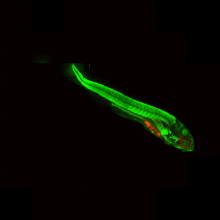
Normalize the orientation of the images of the Zebrafish embryos.

In the documentation webpage, the aim of the workflow is to normalize the orientation of the images of the Zebrafish embryos, find the point of injection of tumor cells and measure the distribution of Cy3 stained tumor foci.

ImageJ macro implementation of the Workflow described in Ghotra et al (2012). Note that currently only the angle and orientation normalization is implemented in this version.

Sample images are linked in the documentation webpage. 

has function
Description

ImageJ plugin to analyze changes in vessel diameters, described in Fernández er al (2014). More specifically the paper describes the measurement of isolated retinal arterioles (ca 50 micrometer diameter) but can be used for diameter measurements of similar vessel structures.

Description

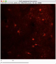
The workflow measures the growth of cells in 3D, combining an ImageJ macro for preprocessing and successive tracking using Imaris.  

The sample dataset (available in the github repository) contains 2-Photon images of neurons. The neurons were imaged in 3D at two time frames.To allow measuring significant differences in cell volume, the time gap between the frames is large (ca. 30 min) and the animal was removed in the waiting phase. For this reason, there is a considerable shift in sample position between the frames that has to be corrected before cell detection and tracking.

The workflow consists of following steps:

1. Import of single tiff slices [imageJ macro]

2. Organizing the data in a 4D time series with 2 time frames [imageJ macro]

3. Correction of shift between the time frames by rigid registration [imagJ macro]

4. Bleaching correction [imageJ macro]

5. Export of preprocessed image data in ics/ids format [imageJ macro]

6. Import of ics/ids data to Imaris [Imaris]

7. Cell object detection as "Imaris Surface Object" [Imaris]

8. Tracking cell objects over time [Imaris]

9. Split Tracks (use Imaris XT extension "Split Tracks") to generate single cell objects [Imaris]

10. Export the statistics: Select the complete folder, go to the statistics tab and use ‚Full Export’ [Imaris]

The preprocessing macro can be referenced here.

The sample images were acquired by Cordula Ulbrich (Petzold Group at German Center of Neurodegenerative Disesases (DZNE), Bonn, Germany).

Input data type: tiff

Output data type: data table

has function
Description
Tracer allows the user to create a trace along a structure in an image. It uses the underlying molecule positions, not the rendered image.
need a thumbnail
Description
Imaris is a commercial 3D image visualisation and analysis tool. It can be used to produce complex 3D animations that include multiple volume and surface elements in several channels, as well as clipping planes and annotations such as text and arrows. Movies interpolate seamlessly between user-defined key frames, and properties such as viewing angle, zoom and visibility of each element can be changed during the animation. These features allow effective communication of results based on image data.
need a thumbnail