Contents
| Image | Title | Category | Type | Description | Updated |
|---|---|---|---|---|---|
|
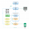
Render | Software | Collection | A collection of Java tools and HTTP services (APIs) for rendering transformed image tiles that includes: |
03/16/2022 - 07:15 |
|
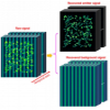
EVER-ImageJ-Plugin | Software | Component | Removal of heterogeneous background from image data of single-molecule localization microscopy, using extreme value-based emitter recovery (EVER). Quote:
|
04/25/2023 - 19:45 |
|
pyTFM | Software | Collection | 10/18/2021 - 08:40 | |
|
napari-pyclesperanto-assistant | Software | Collection, Component | The napari-pyclesperanto-assistant is a yet experimental napari plugin for building GPU-accelerated image processing workflows targeting life-sciences and bio-image analysis. It is part of the clEsperanto project. It uses pyclesperanto and pyopencl as backend for processing images. |
07/10/2021 - 13:03 |
| Lecture Bio-image analysis, biostatistics, programming and machine learning for computational biology at the Biotechnology Center, TU Dresden, 2021 | Training Material | Thie lecture is for Python beginners who want to dive into image processing with Python. It specifically aims for students and scientists working with microscopy images in the life sciences. We start with python basics, dive into descriptive statistics for working with measurements and matplotlib for plotting results. |
07/10/2021 - 12:14 | ||
| Lecture Applied Bio-image Analysis at Biotechnology Center, TU Dresden, 2020 | Training Material | 07/10/2021 - 12:10 | |||
| Customizing ImageJ | Training Material | These slides give an introduction to user interfacre customization in ImageJ using ImageJ Macro and to ImageJ Macro Markdown. |
07/10/2021 - 12:05 | ||
| Sharing and licensing material | Training Material | Talk about sharing and licensing materials such as [raw] data, manuscripts, code, slides, ... at the EMBO Course on Advanced Methods for Bio-image Analysis 2021 virtually in Heidelberg |
07/10/2021 - 12:02 | ||
| On-the-fly image processing with Python and napari | Training Material | These slides were shown in the "On-the-fly image processing session with Python and napari" session by Robert Haase of the Smart Microscopy Workshop at the Center for Cellular imaging at the University of Gothenbourg in May 2021. |
07/10/2021 - 11:57 | ||
|
AnnotatorJ | Software | Component | AnnotatorJ is a Fiji Plugin to ease annotation of images, particulrly useful for Deep Learning or to validate an alogorithm. Interestingly, it allows annotation for instance segmentation, semantic segmentation, or bounding box annotations. It includes toolssuch as active contours to ease these annotations. |
07/09/2021 - 10:03 |
|
NDPI Export Regions Tool | Software | Workflow | The tool exports rectangular regions, defined with the Click the button and select the input folder. The input folder must contain pairs of ndpi and ndpa files. The regions will be exported to a subfolder of the input folder names |
05/24/2023 - 13:55 |
| |
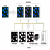
APEER | Software | Collection, Component | Machine Learning made easy APEER ML provides an easy way to train your own machine learning
|
07/01/2021 - 12:52 |
| Image processing with Python | Training Material | This lesson shows how to use Python and skimage to do basic image processing. |
06/30/2021 - 15:51 | ||
| Image Analysis Training Resources | Training Material | This is a resource for image analysis training material, with a focus on research in the life sciences. Currently, this resource is mainly meant to serve image analysis trainers, helping them to design courses. However, we might add more text (or videos) to the material such that it could also be used by students for self-directed study. |
06/30/2021 - 14:24 | ||
|
Cell Tracking Challenge Dataset | Dataset | The datasets consist of ANNOTATED 2D and 3D time-lapse video sequences of fluorescent counterstained nuclei or cells moving on top or immersed in a substrate, along with 2D Bright Field, Phase Contrast, and Differential Interference Contrast (DIC) microscopy videos of cells moving on a flat substrate. The videos cover a wide range of cell types and quality (spatial and temporal resolution, noise levels etc.) In addition, we provide 2D and 3D videos of synthetic fluorescently stained cells and nuclei of different shapes and motion patterns. |
06/30/2021 - 12:33 | |

|
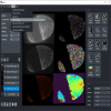
Phindr3D | Software | Workflow | Phindr3D is a comprehensive shallow-learning framework for automated quantitative phenotyping of three-dimensional (3D) high content screening image data using unsupervised data-driven voxel-based feature learning, which enables computationally facile classification, clustering and data visualization. Please see our GitHub page and the original publication for details. |
05/26/2021 - 22:30 |
|
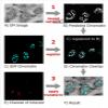
DeepCLEM | Software | Component, Workflow | This Fiji plugin is a python script for CLEM registration using deep learning, but it could be applied in principle to other modalities. The pretrained model was learned on chromatin SEM images and fluorescent staining, but a script is also provided to train an new model, based on CSBDeep. The registration is then performed as a feature based registration, using register virtual stack plugin (which extract features and then perform RANSAc. Editing the script in python gives access to more option (such as the transformation model to be used, similarity by default. |
05/19/2021 - 19:52 |

|
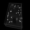
Analyze Spheroid Cell Invasion In 3D Matrix | Software | Workflow | The tool allows to measure the area of the invading spheroïd in a 3D cell invasion assay. It can also count and measure the area of the nuclei within the spheroïd. |
04/29/2023 - 22:40 |
|
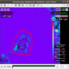
Analyze Complex Roots Tool | Software | Workflow | This tool allows to analyze morphological characteristics of complex roots. While for young roots the root system architecture can be analyzed automatically, this is often not possible for more developed roots. The tool is inspired by the Sholl analysis used in neuronal studies. The tool creates a binary mask and the Euclidean Distance Transform from the input image. It then allows to draw concentric circles around a base point and to extract measures on or within the circles. |
05/24/2023 - 14:02 |

|
Local Z Projector | Software | Component | Local Z Projector is an ImageJ2 plugin, available in Fiji, that can perform local-Z projection of a 3D stack, possibly over time, possibly very large. LZP performs projection of a surface of interest on a 2D plane from a 3D image. It is a simple tool that focuses on usability and is designed to be adaptable to many different use cases and image quality. |
05/05/2021 - 16:12 |