
Contents
| Image | Title | Category | Type | Description | Updated |
|---|---|---|---|---|---|
|
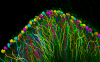
Neurolucida | Software | Collection | Neurolucida is a powerful tool for creating and analyzing realistic, meaningful, and quantifiable neuron reconstructions from microscope images. Perform detailed morphometric analysis of neurons, such as quantifying 1) the number of dendrites, axons, nodes, synapses, and spines, 2) the length, width, and volume of dendrites and axons, 3) the area and volume of the soma, and 4) the complexity and extension of neurons. See 10.3389/fnins.2012.00049 |
03/05/2020 - 11:28 |
|
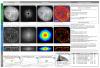
SIMcheck | Software | Collection | SIMcheck is an ImageJ plugin suite for assessing the quality and reliability of Structured Illumination Microscopy (SIM) data. The quality of the raw data, the quality of the reconstruction and the calibration of the microscope can be tested. |
02/05/2019 - 12:18 |
|
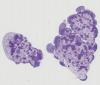
Pyxit segmentation model prediction | Software | Component | This is the "prediction step" of the Pyxit segmentation model builder. It is a learnable segmentation algorithm based on ground-truth images and segmentation mask. It learns a multiple output pixel classification algorithm. It downloads from Cytomine-Core annotation images+alphamasks from project(s), build a segmentation (pixel classifier) model which is saved locally. Typical application: tumor detection in tissues in histology slides. |
10/18/2019 - 18:49 |
|
Pyxit segmentation model builder | Software | Component | This is a learnable segmentation algorithm based on ground-truth images and segmentation mask. It learns a multiple output pixel classification algorithm. It downloads from Cytomine-Core annotation images+alphamasks from project(s), build a segmentation (pixel classifier) model which is saved locally. Typical application: tumor detection in tissues in histology slides. |
10/18/2019 - 18:52 |

|
Colormine | Software | Collection | Nuget package for conversion between color spaces and calculation of color differences. Color spaces available: -CMY -CMYK -HSL -HSB -HSV -CIE L*a*b* -Hunter LAB -L*C*h* -L*u*v* -RGB -XYZ -YXY Color differences available: -CIE76 -CMC l:c -CIE94 -CIE2000. Online example at http://colormine.org/color-converter |
03/05/2020 - 11:37 |
|
|
Pyxit object classification prediction | Software | Component | This module is for applying classification models on objects. It downloads from Cytomine-Core annotation images and coordinate of annotated objects from project(s) and build a annotation classification model which is saved locally. It downloads from Cytomine-Core annotations images from an image (e.g. detected by an object finder), apply a classification model (previously saved locally), and uploads to Cytomine-Core annotation terms (in a userjob layer). |
10/18/2019 - 18:55 |
|
|
Pyxit object classification model builder | Software | Component |
|
05/02/2023 - 18:27 |
|
SLDC (Segment Locate Dispatch Classify) | Software | Collection | SLDC is an open-source Python workflow. SLDC stands for Segment Locate Dispatch Classify. This framework aims at facilitating the development of algorithms for detecting objects in multi-gigapixel images. Particularly, it provides algorithm developers with a structure to define problem-dependent components of their processing workflow (i.e. segmentation and classification) in a concise way. Every other concern such as parallelization and large image handling are encapsulated by the framework. |
04/28/2023 - 15:08 |
| BioImage Informatics Index (BIII) | Basic page | 09/22/2021 - 19:17 | |||

|
ec-clem autofinder | Software | Component | Automatic registration in 2D or 3D based on detection or binary mask. Takes images with detections already done on it. |
05/31/2023 - 14:09 |
|
|
Icy Protocols | Software | Component | 10/18/2019 - 13:59 | |

|
eC-CLEM | Software | Component | This plugin allows to compute a similarity (translation/rotation/scaling and flipping) transform from pair of points. It is updating the transformed image interactively such that the user get immediate feedback. The transformation is saved and can be applied to any other stack/image. Non rigid deformation can also be applied in 2D or 3D. 3D/3D,2D/3D or 3D /2D can be handled . 3D ROI are enabled, and can be checked with the 3D vtk view (size of ROI can be changed using the ROI stroke width). Some prealignment by rotating in 3D the volume is possible. |
05/31/2023 - 14:10 |
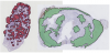
| Quantitative Evaluation of Multicellular Movements in Drosophila Embryo | Training Material | In this chapter you will learn to track cell movements within the monolayer epithelium of a Drosophila embryo.The segmentation of the cells is performed by an ImageJ macro on microscope time lapses (movies) of the embryo presented in the maximum intensity projection (step1). Another macro can optionally be used to discard weak cell–cell junctions that are likely to be segmentation errors(step2). Thet racking is then performed in Matlab from this binary stack (step3). |
01/26/2017 - 15:50 | ||
| About BIII | Basic page | Purpose:Many tools for BioImage Analysis are already available but information about these tools is non-uniform and often focuses on technicalities about the methods implemented rather than the problems the tools can actually solve. Since bioimage analysts focus on applied problems, this information is often inadequate. |
05/17/2023 - 09:13 | ||
|
|
imageHTS | Software | Collection | imageHTS is an R/Bioconductor package dedicated to the analysis of high-throughput microscopy-based screens. The package provides a modular and extensible framework to segment cells, extract quantitative cell features, predict cell types and browse screen data through web interfaces. Designed to operate in distributed environments, imageHTS provides a standardized access to remote data and facilitates the dissemination of high-throughput microscopy-based datasets. | 09/13/2017 - 12:16 |
|
jicbioimage | Software | Collection | The jicbioimage Python package makes it easy to explore microscopy data in a programmatic fashion (python). Exploring images via coding means that the exploratory work becomes recorded and reproducible. Furthermore, it makes it easier to convert the exploratory work into (semi) automated analysis work flows. Features:
|
08/17/2018 - 15:27 |
|
|
TurtleSeg: 3D Image Segmentation Software | Software | Collection | TurtleSeg is an interactive 3D image segmentation tool. TurtleSeg has an automated system, Spotlight, for automatically directing the user towards the next steps. Typically, a user loads a 3D image and then manually contour a sparse number of slices, the full 3D segmentation can then be built automatically. |
11/13/2019 - 12:58 |
|
|
Huygens Remote Manager | Software | Collection | The Huygens Remote Manager is an open-source, efficient, multi-user web-based interface to the Huygens software by Scientific Volume Imaging for parallel batch deconvolutions. |
03/05/2020 - 11:43 |
|
|
IceImarisConnector | Software | Collection | IceImarisConnector is a simple commodity class that eases communication between Bitplane Imaris and MATLAB or python using the Imaris XT interface. | 09/13/2017 - 12:16 |
|
|
XuvTools | Software | Collection | XuvTools (pronounced ex-you-vee-tools) is a fully automated 3D stitching software for biomedical image data, typically confocal microscopy images. XuvTools runs on Microsoft Windows XP and Vista, Linux and Apple Mac computers. It supports 32 and 64bit operating systems (with 64bit highly preferred). XuvTools is free and open source software (see Licensing), so you can start using it immediately. Go to Downloads and give it a try. |
03/05/2020 - 11:48 |