
Contents
| Image | Title | Category | Type | Description | Updated |
|---|---|---|---|---|---|
|
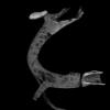
Tube un-winder | Software | Component | This macro can be used to un-wide a tubular structure and flatten its surface (like peeling of and flattening the skin of a banana). The macro can only process a single channel 3D stack but it is easy to process multiple channels by exporting and importing ROI manager selections. Technically the macro computes the radial average intensity projection inside a ring centred on the radial symmetry axis of the object. The final image is a radial mapping of the intensity (radial angle along X, axial length along Y). The example image is available in the documentation link. |
04/28/2023 - 16:05 |
|
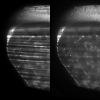
SPIM de-striper | Software | Component | This macro implements a filter that is meant to attenuate close to parallel intensity stripes in an image, such as often happening in light sheet microscopy. The results are usually decent even when the stripes show a large angular spread due to light sheet refraction at the sample surface. The filter can process a 3D stack but the processing is performed slice by slice. Example image is available in the documentation link. |
04/28/2023 - 16:22 |

|
Morphological Segmentation (ImageJ) | Software | Component | Morphological Segmentation is an ImageJ/Fiji plugin that combines morphological operations, such as extended minima and morphological gradient, with watershed flooding algorithms to segment grayscale images of any type (8, 16 and 32-bit) in 2D and 3D. Morphological Segmentation runs on any open grayscale image, single 2D image or (3D) stack. If no image is open when calling the plugin, an Open dialog will pop up. The user can pan, zoom in and out, or scroll between slices (if the input image is a stack) in the main canvas as if it were any other ImageJ window. |
03/02/2020 - 21:05 |
| |
Tracking of focal adhesions in 2D time lapse movies | Software | Workflow | Tracking of focal adhesions includes a number of challenges: |
05/03/2023 - 16:30 |

|
Tissue analysis from histological sections | Software | Workflow | This macro batch processes all the 2D images (tif and jpg files) located in a user defined folder by calling Fiji Weka trainable segmentation to classify each pixel, and reports the areas of each class in a human readable results table. The classifier to be applied to each image should be previously trained on a representative image by an expert and exported to file (Save classifier) into the image folder to be processed. |
04/28/2023 - 16:08 |
|
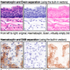
Quantifying staining in tissue sections | Software | Component | [no download link, this description itself explains the steps to quantify staining in tissue sections] The Color Deconvolution plugin for ImageJ can be used to digitally separate up to three stains from brightfield images, after which standard ImageJ commands can be used. The algorithm is described in Ruifork and Johnston (2001). **However**, it is **very** important to take into consideration the caveats on the linked URL. |
04/29/2023 - 22:32 |

|
Blood vessel segmentation and network analysis | Software | Workflow | This macro segments blood vessels in a 3D stack. It is suited for well-contrasted images (low background) and works better if the width of the vessels of interest is reasonably uniform.
Sample image: 1 sample image: 2 |
04/28/2023 - 16:16 |
| |
Volocity - 3D Object Segmentation and Tracking | Software | Component | In the commercial image analysis software "Volocity", automated measurement protocols can be constructed by dragging, dropping and configuring a sequence of individual "tasks". By combining the "Find Objects" task with a subsequent "Track" task, 3D objects can be identified and followed over time. The initial "Find Objects" segmentation can be refined, e.g. using "Separate Touching Objects"; and tracking results in the form of "Measurement Items" can be viewed in tabular form, as a graph, etc. |
05/24/2018 - 01:31 |
|
|
Autofocus | Software | Component | Autofocus hyperstack macro: Select the in focus frame from each slice of a hyperstack and create a new stack of just the in focus frames. Based on algorithm F-11 "Normalized Variance". Original macro by Andy Weller. |
05/03/2023 - 17:32 |
|
KymographTracker | Software | Component | Generation of Kymographs using 2D+t images. In the generated kymographs, objects can be tracked and the results are visualized. |
10/29/2019 - 18:25 |

|
Protein Micro-array Analysis | Software | Workflow | This macro performs measurements of average and standard deviation intensity inside wells of a protein microarray (the number of wells is limited to 250, the image should be cropped for larger arrays). |
04/28/2023 - 16:12 |
|
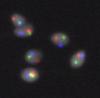
FISH signals detection | Software | Component | The macro segments and classifies human spermatozoids nuclei (DAPI) based on the number of FISH signals (spots) they contain. It reports the percentage of occurrences of user defined classes (combinations of spot multiplicity in the FISH channels) as well as the position (point selections) of the detected nuclei falling in these classes. The input image should be an hyperstack with 4 channels: DAPI (first channel) and three FISH channels. The images are typically obtained as a maximum intensity projection of few channels (confocal) or a single z slice acquisition (widefield). |
04/28/2023 - 16:03 |
|
|
Aphelion | Software | Collection | A commercial image analysis software. It's interface allows to easily perform measurements and image analysis. Your actions can be recorded and a macro (in a basic script language) can then be created. Almost no knowledge in programming is needed. You can also use python. A SDK is also available to develop stand alone applications in c++. Additional modules allow to use specific operations (3D operators... |
03/02/2020 - 21:51 |

|
CellProfiler Examples - Speckle Counting | Software | Component | Quote:
|
04/29/2023 - 14:55 |
|
Quantitative analysis of focal adhesion dynamics | Software | Workflow | 05/16/2018 - 23:31 | |
|
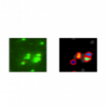
CellProfiler Examples - Colocalization | Software | Workflow | Quote:
|
04/29/2023 - 14:22 |
| |
Microtubule end tracking in Drosophila Oocyte | Software | Workflow | Microtubule end tracking in live cell fluorescent images of Drosophila oocyte involves overcoming the following challenges, which can be tackled by a series of preprocessing steps and tracking described in Parton et al (2011) |
05/03/2023 - 10:51 |

|
Quantitative analysis of focal adhesions | Software | Workflow | Simple workflow description for ImageJ, step-by-step description for delineating focal adhesions, count and characterize their positions. Measurement of dynamics is not involved. |
05/25/2018 - 18:48 |
|
|
3D cell tracking and quantification of shape changes | Software | Workflow | The workflow includes segmentation, tracking and quantifying morphological dynamics of moving cells in 3D. The authors have implemented the workflow in Matlab, but so far there is no download link provided. To apply this workflow, we recommend to contact the authors or to implement the worflow based on the detailed description in the original paper. |
05/31/2018 - 22:10 |
|
Multi Kymograph | Software | Collection | This macro and plugins suite for ImageJ (and Fiji) serves to measure the velocity of moving structures and visualize them, from image time series (2D over time). The module can be installed in ImageJ as a Macro Menu and each function/component can be called separately. The full workflow consists in calling some, or all, the functions sequentially in order to get from the image preparation (e.g. filtering and visualization of tracks) to the production of the kymographs (time vs. distance plot) and their analysis (retrieving the velocities). |
05/03/2023 - 11:09 |