
Contents
| Image | Title | Category | Type | Description | Updated |
|---|---|---|---|---|---|
| Introduction to R for bioimage analysis | Training Material | This session covers the basics of the R programming language in the context of a bioimage analysis workflow. It aims at getting started with the analysis of data extracted from one or several images, here tracking data. Tracking data were extracted using the Fiji plugin TrackMate and are available in |
09/10/2019 - 22:17 | ||
| Deep Learning from scratch | Training Material | Make your first experience with TensorFlow-Keras Part 1 : https://canal-ua.univ-angers.fr/avc/courseaccess?id=4797
Part 2 : https://canal-ua.univ-angers.fr/avc/courseaccess?id=4796
Part 3 : https://canal-ua.univ-angers.fr/avc/courseaccess?id=4809
|
08/29/2019 - 10:22 | ||

|
FAIRsharing Euro-BioImaging collection | Dataset | FAIRsharing is a registry of standards, including deposition databases and data repositories. The collection gathered by Euro-BioImaging contains a list of public databases with bioimaging data sets. |
03/10/2023 - 23:56 | |
|
DeepCell | Software | Component |
DeepCell is neural network library for single cell analysis, written in Python and built using TensorFlow and Keras. DeepCell aids in biological analysis by automatically segmenting and classifying cells in optical microscopy images. This framework consumes raw images and provides uniquely annotated files as an output. The jupyter session in the read docs are broken, but the one from the GitHub are functional (see usage example ) |
10/19/2020 - 17:09 |
|
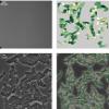
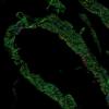
muscle cross-sections | Dataset | Muscle cross-section with immunofluorescence , to count myofibers |
08/26/2019 - 11:59 | |
|
Mouse embryos | Dataset | Fluorescence microscopy cannot be used to image human embryos to determine embryo viability for in vitro fertilization because the introduction of exogenous fluorescent dyes is considered a toxic procedure. As a result, embryo viability has been measured primarily using differential interference contrast (DIC). |
08/26/2019 - 11:49 | |
|
two-photon images of dendritic spines | Dataset | Raw data of three types of dendritic spines imaged in biphoton (mushroom, tubby, thin spines). Hippocampal neurons from mouse organotypic slice cultures
If you use this dataset , please cite M. U. Ghani, F. Mesadi, S. D. Kanik, A. O. Argunsah, A. Hobbiss, I. Israely, D. Unay, T. Tasdizen, and M. Cetin, "Shape and appearance features based dendritic spine classification," Journal of Neuroscience Methods. |
08/26/2019 - 11:45 | |
|
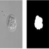
Human colon tissue | Dataset | One of the principal challenges in counting or segmenting cells or cell nuclei is dealing with clustered objects such as in tissues. To help assess algorithms' performance in this regard, synthetic 3D image sets of human colon tissue are provided in two diferent levels of quality: high SNR and low SNR. Ground truth is available as well. |
08/26/2019 - 11:33 | |
|
Clustered Cell Nuclei Data | Dataset | One of the principal challenges in counting or segmenting cells or cell nuclei is dealing with clustered objects. To help assess algorithms' performance in this regard, synthetic 3D image sets of HL60 cell line are provided consisting of four subsets with increasing degree of clustering. Each subset is also provided in two diferent levels of quality: high SNR and low SNR. Ground truth is available as well. |
08/26/2019 - 11:29 | |
|
DragonFly | Software | Collection | Dragonfly is a software platform for the intuitive inspection of multi-scale multi-modality image data. Its user-friendly experience translates into powerful quantitative findings with high-impact visuals, driven by nuanced easy-to-learn controls. For segmentation: It provides an engine fior machine Learning, Watershed and superpixel methods, support histological data . It offers a 3D viewer, and python scripting capacities . It is free for reserach use, but not for commercial usage. |
10/17/2019 - 12:18 |
|
CSIRO science image library | Dataset | collection of real life science images and videos from Australian research labs. including photographs of plants, microscopy equipments, microscopy images. |
10/28/2025 - 01:58 | |
|
Neuroglancer | Software | Component | Web based viewer developped for google for very big data: Neuroglancer is a WebGL-based viewer for volumetric data. It is capable of displaying arbitrary (non axis-aligned) cross-sectional views of volumetric data, as well as 3-D meshes and line-segment based models (skeletons). The segmentation has to be done before loading the dataset, it is not done Inside the viewer. This is not an official Google product. It has among other the nice feature of beeing able to generate url for sharing a specific view. Note that the only supported browser for now are |
04/27/2023 - 13:00 |

|
Breast Cancer Histopathological Database (BreakHis) | Dataset | 2,480 benign and 5,429 malignant annotated histophatology dataset of cancer breast tissue from 82 patients using different magnifying factors (40X, 100X, 200X, and 400X) |
07/26/2019 - 16:25 | |
|
3D Segmentation & visualization of nanoparticles attached to the surface of red blood cells | Software | Workflow | 05/24/2023 - 16:47 | |

|
Using ImageJ to Analyze the Size, Shape, and Directionality of Networks of Coupled Astrocytes | Software | Workflow | The research goal of this paper was to provide unbiased counts of labeled astrocytes and to estimate the area they cover, further to develop tools for defining the orientation of coupling within astrocyte networks under different stimuli. |
05/24/2023 - 17:28 |
|
WormScan | Software | Collection | 10/15/2019 - 10:59 | |

|
Ikosa | Software | Collection | online image data management system which supports authenticated image upload, cloud-based storage, project-based management and viewing of standard and whole slide images. One can use different annotation tools to highlight important objects or areas within images. It is the first basic version and new features such as sharing for easy collaboration with your colleagues or first automated analysis applications based on artificial intelligence will be added soon. Ikosa Portal: multi user image data management |
10/16/2019 - 11:13 |
|
Protein Array Analyzer for ImageJ | Software | Collection | Protein array is used to analyze protein expressions by screening simultaneously several protein-molecule interactions such as protein-protein and protein-DNA interactions. In most cases, the detection of interactions leads to an image containing numerous lines of spots that will be analyzed by comparing tables of intensity values. |
10/19/2020 - 17:09 |
|
|
Zebrafish larvae - Widefiled/Brightfield | Dataset | Datasets originate from internal test runs at ACQUIFER on the Imaging Machine (see also application note on "The ACQUIFER PlateViewer: A tool for visualizing high content screening data and supervised feedback microscopy") |
04/29/2019 - 12:22 | |
|
Medaka embryo in 96 well plate - Widefield Brightfield | Dataset | 102 hpf medaka embryos in 96 well plate (4 embryo/well) - brightfield - 2X magnification - ACQUIFER Imaging Machine Gierten, Jakob, et al. "Automated high-throughput heart rate measurement in medaka and zebrafish embryos under physiological conditions." bioRxiv (2019): 548594. Used as benchmark dataset for Multi-Template-Matching by Thomas and Gehrig See implementation in Fiji https://github.com/LauLauThom/MultipleTemplateMatching |
04/29/2019 - 12:20 |