
Contents
| Image | Title | Category | Type | Description | Updated |
|---|---|---|---|---|---|
|
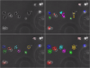
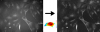
AVEMAP | Software | Component | Measures wound-healing assay videos, For each video, the velocity and the order parameter are analyzed in time and space to extract quantitative parameters characterizing the cell motility phenotype. The different conditions (videos) can then be classified according to these parameters. |
03/10/2023 - 23:13 |

|
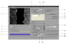
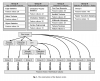
NeuronMetrics | Software | Collection | The invention comprises a software tool, NeuronMetrics, which functions as a set of modules that run in the open-source program ImageJ. NeuronMetrics features a novel method for estimating neural “branch number” (a measure of the axonal complexity) from two-dimensional images. In addition, the tool features a novel method for modeling neural structure in large “gaps” that result from image artifacts. |
01/29/2018 - 09:49 |
|
|
Hessian by Philippe Thévenaz | Software | Component | Computes image Hessian Splines: A Perfect Fit for Signal and Image Processing |
09/12/2017 - 17:53 |
|
|
Laplacian by Philippe Thévenaz | Software | Component | Computes image Laplacian
Based on the algorithm described in the paper below. Splines: A Perfect Fit for Signal and Image Processing |
09/12/2017 - 17:48 |
|
|
Matlab compiler runtime | Software | 09/12/2017 - 17:00 | ||
|
|
gradient by Philippe Thévenaz | Software | Component | Computes image gradient
Based on the algorithm below. Splines: A Perfect Fit for Signal and Image Processing |
09/12/2017 - 17:14 |
|
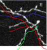
Neural Circuit Tracer | Software | Collection | Neural Circuit Tracer (NCTracer) is open source software for automated and manual tracing of neurites from light microscopy stacks of images. NCTracer has more than one workflow available for neuron tracing. |
04/11/2018 - 12:01 |
| |
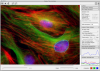
Microscopy Image Browser (MIB) | Software | Collection | Microscopy Image Browser (MIB) is a high-performance Matlab-based software package for advanced image processing, segmentation and visualization of multi-dimensional (2D-4D) light and electron microscopy datasets. |
05/12/2023 - 17:52 |
|
AnaMorf | Software | Component | AnaMorf is a plug-in developed for the ImageJ platform to analyse the microscopic morphology of filamentous microbes. The program returns average data on a population of mycelial elements, using the descriptors projected area, circularity, total hyphal length, number of hyphal tips, hyphal growth unit, lacunarity and fractal dimension. The plug-in accepts as input a user-specified directory of images, analysing each and outputing tabulated results. |
10/30/2025 - 04:59 |
|
Kappa | Software | Component | Kappa is a Fiji plugin for Curvature Analysis. It allows a user to measure curvature in images in a convenient way. You can trace an initial shape with a B-Spline curve in just a few clicks and then fit that curve to image data with a minimization algorithm. It’s fast and robust. |
09/21/2017 - 16:50 |
| Kota Miura (ed) "Bioimage Data Analysis", Textbook, Wiley | Training Material | Bioimage Data Analysis provides detailed explanations and example workflows of combining various image processing algorithms to construct practical workflows using scripting. http://www.imaging-git.com/olympus-website-bioimage-data-analysis https://www.researchgate.net/publication/297738620_Bioimage_Data_Analysis |
08/03/2020 - 14:50 | ||
|
iTrack4U | Software | Component |
Please cite Cordeliéres et. al. (2013) when using this software package! |
05/21/2018 - 01:22 |
|
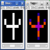
ImageJ Analyze Skeleton | Software | Component | This plugin tags all pixel/voxels in a skeleton image and then counts all its junctions, triple and quadruple points and branches, and measures their average and maximum length. Tags are shown in a new window displaying every tag in a different color. You can find it under [Plugins>Skeleton>Analyze Skeleton (2D/3D)]. See Skeletonize3D for an example of how to produce skeleton images. |
04/27/2023 - 14:09 |
|
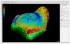
MorphoGraphX | Software | Collection | MorphoGraphX is a free Linux application for the visualization and analysis of 3D biological datasets. Developed by researchers, it is primarily used for the analysis and quantification of 3D live-imaged confocal data sets. The main research interests adressed by MorphoGraphX are:
|
03/03/2020 - 20:54 |
| Rafael C. Gonzalez, Richard E. Woods."Digital Image Processing", Pearson, 2008 | Training Material | From the publisher: "Completely self-contained–and heavily illustrated–this introduction to basic concepts and methodologies for digital image processing is written at a level that truly is suitable for seniors and first-year graduate students in almost any technical discipline. |
09/12/2017 - 11:58 | ||
|
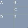
FigureJ | Software | Component | This plugin achieves easy creation of image figures for publications, reports, projects.
|
04/29/2023 - 11:34 |
|
|
hIPNAT | Software | Collection | hIPNAT (hIPNAT: Image Processing for NeuroAnatomy and Tree-like structures) is a set of tools for the analysis of images of neurons and other tree-like morphologies. It is written for ImageJ, the de facto standard in scientific image processing. It is available through the ImageJ Neuroanatomy update site. |
03/03/2020 - 17:55 |
|
CIDRE | Software | Collection | CIDRE is a retrospective illumination correction method for optical microscopy. It is designed to correct collections of images by building a model of the illumination distortion directly from the image data. Larger image collections provide more robust corrections. Details of the method are described in K. Smith, Y. Li, F. Ficcinini, G. Csucs, A. Bevilacqua, and P. Horvath |
04/29/2023 - 14:27 |
|
wnd-charm | Software | WND-CHARM is a multi-purpose image classifier that can be applied to a wide variety of image classification tasks without modifications or fine-tuning, and yet provides classification accuracy comparable to state-of-the-art task-specific image classifiers. WND-CHARM can extract up to ~3,000 generic image descriptors (features) including polynomial decompositions, high contrast features, pixel statistics, and textures. These features are derived from the raw image, transforms of the image, and compound transforms of the image (transforms of transforms). |
10/18/2018 - 17:35 | |

|
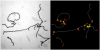
Rivulet | Software | Component | "we propose a novel automatic 3D neuron reconstruction algorithm, named Rivulet, which is based on the multi-stencils fast-marching and iterative back-tracking. The proposed Rivulet algorithm is capable of tracing discontinuous areas without being interrupted by densely distributed noises." This plugin can be used with default parameters or with user-defined parameters. Example image obtained from Rivulet Wiki website (https://github.com/RivuletStudio/Rivulet-Neuron-Tracing-Toolbox/wiki) |
04/28/2023 - 15:16 |